Bowel Examination (Tests, Scans, Scopes) Pictures, Diagnosis
Digestive problems are among the most common medical complaints. It can be acute where the symptoms last for a short period of time and resolve with/without treatment, or it can be chronic where the symptoms persist or occur episodically. Although the symptoms along with a case history may be sufficient for a diagnosis, at times additional diagnostic investigation may be necessary. Theses tests, scans and scopes are intended to identify the underlying problem in order to reach an accurate diagnosis and determine the most appropriate treatment.
Bowel Diagnosis
Not all digestive problems may be bowel problems. The gastrointestinal tract, or gut for short, starts at the mouth and ends at the anus. Food is consumed, digested and absorbed in the bowels, with waste substances being expelled as stool. But there are other organs that play an integral role in the functioning of the digestive system as a whole. This includes the liver, pancreas and gallbladder.
The human gut is about 9 to 10 meters (29 to 32 feet) in length with the small intestines comprising about 6 meters (approximately 20 feet) and the large intestine being about 1.5 meters (around 5 feet). Most of the digestive organs are contained within the abdominal cavity. The coiling of the intestines within the abdomen allows for its length to be compactly held within this cavity.
The length and complexity of the gastrointestinal tract (gut) means that a problem can arise anywhere and present with often similar symptoms. It can be as vague as abdominal pain, nausea and vomiting, alterations in bowel movement (diarrhea and constipation) and as generalized as malaise (feeling unwell), changes in appetite and unintentional weight loss.
Without further diagnostic investigation, it can sometimes be difficult diagnosing the underlying problem just by the symptoms alone.
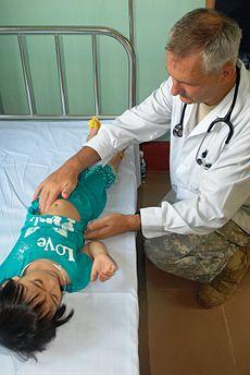
Physical Examination
After taking down your case history and isolating the most relevant symptoms, a doctor will then conduct a physical examination. With regards to the bowels, the following techniques may be employed to isolate a problem:
- Inspection – visual examination of the abdomen to note any protrusions, swelling or discoloration among other abnormalities.
- Palpation – feeling the abdomen in quadrants by hand with deeper palpation for areas where pain or other abnormalities have been noted.
- Percussion – tapping of the abdomen to identify different organs by sound as well as noting shifting dullness when there is fluid accumulation.
- Auscultation – where abdominal sounds are heard through the use of a stethoscope.
- Throat and neck examination – both externally and internally.
- Rectal examination – with or without a speculum.
Based on the case history, clinical presentation and examination findings, a doctor may decide that further diagnostic investigation is necessary.
Bowel Tests
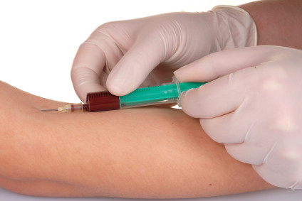
Blood
There are a range of blood tests that may be performed but most will not specifically isolate a problem to the bowel. The results of this test could indicate an abnormality but the problem may lie with the liver or pancreas rather than the bowel specifically. Blood tests may reveal anemia associated with blood loss, antibodies to bowel microbes and markers of inflammation among other biochemical abnormalities.
Stool
Stool tests are therefore more accurate as it is able to assess the intestinal contents for any abnormalities. Some of the stool tests may be used to identify:
- Nutrients that are partially digested or not digested at all.
- Blood in the stool, even trace amounts. Read more on the fecal occult blood test (FOBT).
- Eggs and parasites in the stool.
- Bacteria or protozoa in the stool.
- Enzymes in the stool.
- White blood cells (WBCs) in the stool.
- Proteins and antibodies in the stool.
Rea more on stool analysis.
Secretions
Secretory tests collect samples from the gut to analyze the contents. These tests assess the secretions of the gut to aid with the diagnosis of a condition. It may require the administration of certain drugs or hormones to stimulate the relevant secretions.
Bowel Scopes
The development of endoscopic procedures has revolutionized medicine, particularly in gastroenterology. Thin, flexible tubes with a camera and light at the end can be used to view the interior of the gut. These devices are known as endoscopes. A scope can be inserted through the mouth or anus to examine a portion of the bowel. Scopes may also be inserted through the abdominal wall (laparoscopy) as part of a surgical procedure to examine the abdominal contents including the bowels.
- Upper gastrointestinal (GI) endoscopy where an endoscope is inserted through the mouth to examine the interior of the esophagus, stomach and first part of the small intestine. It is also known as esophagogastroduoscopy.
- Enteroscopy where the small intestine can be viewed. A double balloon endoscopy allows for tiny balloons to be inflated in order to examine a specific section of the small intestine.
- Colonoscopy where the anus, rectum and colon can be viewed. Similar procedures include a sigmoidoscopy which examines up the last part of the colon known as the sigmoid, or proctoscopy to examine the rectum (rectoscopy) and anus (anoscopy).
- Capsule endoscopy is where a pill-shaped camera is swallowed and pictures are taken of the bowel and sent wirelessly to a recorder. There are no attachments to remove the scope and it is eventually passed out of the rectum.
A biopsy may be conducted during endoscopic procedures. Here a small tissue sample is collected and examined to identify any abnormalities.
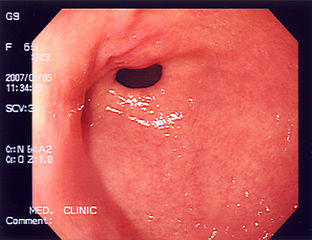
Picture of stomach seen during an endoscopic examination
Bowel Scans
Imaging studies uses x-rays, magnetic and radio waves, and ultrasound waves to form images of the internal structures of the body. Some of these imaging studies require the use of contrast dyes to enhance the visualization of structures.
X-Rays
X-rays uses a form of electromagnetic radiation. A plain x-ray may identify foreign objects and vaguely indicate soft tissue abnormalities but it is mainly bones that are clearly visible on an x-ray. These rays do not pass through denser tissue like bones so a barium meal or barium swallow may be necessary to highlight the soft tissue of the gut on an x-ray.
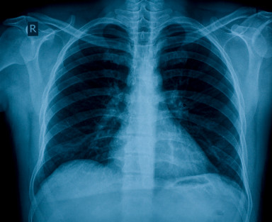
Picture of an x-ray
CT Scan
CT (computerized tomography) scan involves taking x-rays from different angles and combining it to form 3D cross-section images of organs. A radiocontrast dye is usually necessary for a CT scan involving the bowels.
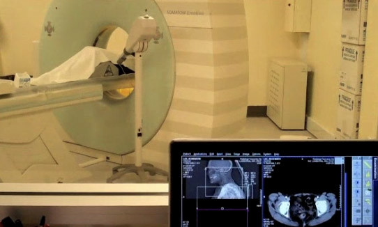
Picture of a CT scan machine and from an actual scan
MRI
MRI (magnetic resonance imaging) uses radio waves and magnetic fields to create detailed cross-sectional images of the body. It can also be adapted to form 3D images.
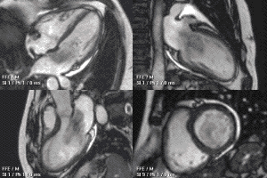
Picture from MRI scan
Ultrasound
Ultrasonography uses ultrasound waves to form images. The emitter produces the waves and the receiver detects waves that bounce back to form images of the target area. It is usually used from outside of the body but some ultrasound devices may be placed within the body for examination.
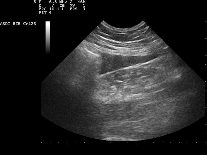
Picture from an abdominal ultrasound


