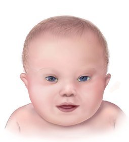
Down Syndrome Babies, Facts, Pictures, Features and Diagnosis
What is Down Syndrome?
Down syndrome, also known as Trisomy 21, is a genetic disorder resulting from possession of an extra copy of chromosome 21. It is named after Dr Langdon Down who first recognized and described the condition in 1866. The genetic origin of Down syndrome was identified much later by scientists in 1959. Individuals with this disorder suffer from varying degrees of learning impairment as well as having typical physical features such as a flat facial profile with upward slanting eyes, short neck, a single palmar crease, and various other recognizable characteristics.
The severity of symptoms in Down syndrome may vary from mild to severe. The condition is often associated with congenital heart disease, acute leukemia, and signs of premature aging. Down syndrome occurs in about 1 in 700 live births. The frequency of Down syndrome seems to rise with increasing maternal age. Screening for Down syndrome is now routinely offered in many developed countries. Genetic counseling is recommended for a person with a family history of Down syndrome who wishes to have a baby.
Genetic Defects in Down Syndrome
Within each nucleus of the human cell there are a set of 46 chromosomes, arranged in 23 pairs. Half of these chromosomes are derived from the mother and half from the father. During gametogenesis, which is the development and production of the male and female germ cells necessary to form a new individual, the number of chromosomes is halved. This ensures that the number of chromosomes remains constant and is not doubled at each conception. The reduction process whereby the chromosome number is halved during gametogenesis is known as meiosis. When fertilization occurs, resulting in conception, 23 chromosomes from the male partner comes together with 23 chromosomes from the female partner. The baby thus conceived has a total of 46 chromosomes.
Trisomy 21
In certain circumstances, however, errors may occur during conception which can give rise to chromosomal abnormalities. If during meiosis the chromosomes of a pair fail to separate then both members of the pair pass into the same gamete (gametes are the male and female germ cells). If this gamete is fertilized by a normal gamete, it will result in a zygote (the fertilized egg) with an additional chromosome. Different chromosomal abnormalities are associated with specific conditions or syndromes.
In Down syndrome or trisomy 21, there is one number 21 chromosome from each partner, but there is also an extra chromosome 21 (which may come from either partner) so that there are three chromosomes 21 instead of two. This will occur in every cell of the body. Genes present in each chromosome are responsible for all characteristics, growth, and development of a human being. The genes carried by the extra chromosome 21 give rise to all the characteristic features of Down syndrome.
Translocation
In a small number of cases, some people with Down syndrome do not inherit an extra chromosome 21. Instead, they only inherit some extra chromosome 21 genes which may be carried by another chromosome. This is known as translocation. In most cases this occurs during conception without any known cause. In some cases, however, a parent may be a balanced carrier of a translocation. This means that the parent’s cells contain 2 copies of chromosome 21 but some of the genes are attached to another chromosome. If a baby inherits this chromosome which contains the extra genes from chromosome 21, he/she will suffer from Down syndrome.
Mosaic Down Syndrome
In another form of Down syndrome known as mosaic Down syndrome, additional genes from chromosome 21 may be inherited but this defect does not occur in all cells of the body. Changed physical characteristics or the degree of mental impairment will vary according to the type and percentage of cells with extra genes from chromosome 21. Mosaic Down syndrome can be differentiated from trisomy 21 by genetic testing.
Risk Factors of Down Syndrome
- Increasing maternal age is the major risk factor for Down syndrome.
- Parents who have once conceived a child with Down syndrome.
- The risk may be 100% if a parent is a carrier of a chromosome 21 translocation.
- Increased risk if either partner has Down syndrome.
Characteristic Features of Down Syndrome
- Flat facial profile.
- Widely spaced and upward slanting eyes.
- Narrow palpebral fissure (the opening for the eyes between the upper and lower eyelids).
- Presence of an epicanthal fold, the skin of the upper eyelid covering the inner corner of the eye.
- Peripheral silver iris spots (Brushfield’s spots) in the eye.
- Flattened nose bridge.
- A small mouth, often with a large protruding tongue.
- High-arched palate.
- Round head.
- Short neck with abundant neck skin.
- Dysplastic or malformed ears.
- Muscle hypotonia or poor muscle tone.
- Short, broad hands with a single, transverse palmar crease.
- Incurving fifth digit.
- Widely spaced first and second toes.
- Dysplastic pelvis on X-ray.
A varying range of mental retardation is common but people with Down syndrome usually have a pleasant personality with a fondness for music.
Associated Problems
- Cognitive impairment or learning disability.
- Low IQ.
- Short stature.
- Congenital heart defects such as atrioventricular septal defect (AVSD), ventricular septal defect (VSD), atrial septal defect (ASD), Fallot’s tetralogy, and patent ductus arteriosus (PDA).
- Gastrointestinal problems such as esophageal and duodenal atresia or stenosis, Hirschsprung disease, and imperforate anus.
- Sight and hearing problems.
- Thyroid problems.
- Acute leukemia.
- Signs of early aging.
- Early-onset Alzheimer’s disease.
Prenatal Screening for Down Syndrome
The American College of Obstetricians and Gynecologists (ACOG) recommends that all pregnant women, regardless of their age, should be offered screening for Down syndrome. Screening should occur before the 20th week of pregnancy. It also advises that all pregnant women, regardless of their age, should have the option of diagnostic testing.
- The triple test – serum AFP, unconjugated estriol, and total human chorionic gonadotropin (hCG) in the mother’s blood are assessed in relation to her age, weight, and duration of pregnancy (as confirmed by ultrasound) to determine the risk of Down syndrome. Free hCG may be a better marker than total hCG.
Quadruple tests additionally measure inhibin-A. Also known as the expanded alpha-fetoprotein (AFP) screening test, this test is usually done between 15 and 20 weeks of pregnancy. An alteration in the AFP and hormone levels in the maternal blood can raise a suspicion of Down syndrome but a normal level will not rule out the diagnosis. - The nuchal translucency test – this is usually done between 11 and 13 weeks of pregnancy. Ultrasound helps in assessing the thickness of the neck skin fold. In Down syndrome, the clear space in the folds of tissue behind the baby’s neck appears larger than normal due to accumulation of fluid. The result when assessed in relation to the mother’s age gives a good amount of accuracy in identifying Down syndrome.
- Other ultrasound screens to look for anatomical defects in the fetus – this is usually done between 18 and 22 weeks of pregnancy. It is mostly recommended for women at high risk of having a baby with Down syndrome.
Prenatal screening can only assess the risk for Down syndrome. These are all non-invasive tests.
Other more invasive tests will be necessary to confirm the diagnosis if Down syndrome is suspected.
Diagnosis of Down Syndrome
Prenatal diagnostic tests for detecting Down syndrome before birth of the baby may increase the chance of miscarriage.
- Amniocentesis is where a small amount of amniotic fluid is removed from the sac surrounding the fetus by means of a thin needle inserted through the abdominal wall under ultrasound guidance. The fluid is examined for chromosomal abnormalities. This test is usually done between 16 and 20 weeks of pregnancy.
- Chorionic villus sampling (CVS) is where a small piece of placental tissue is removed either through the cervix by transcervical catheter, or through the abdomen by transabdominal needle, under ultrasound guidance. This test may be done between 11 and 12 weeks of pregnancy.
- Percutaneous umbilical blood sampling (PUB) is where a needle inserted through the abdominal wall takes a sample of fetal blood from the umbilical cord. This is analyzed for chromosomal abnormalities. This test is usually done after 18 to 20 weeks of pregnancy.
- Fluorescent in situ hybridization (FISH analysis) is a test that can be done on the same sample taken from blood, during amniocentesis, or during CVS. It can determine the number of copies present of a certain chromosome, such as chromosome 21 in Down syndrome.
The above prenatal diagnostic tests may help to detect Down syndrome before birth of the baby but may increase the chance of miscarriage. Karyotyping or chromosome analysis may confirm the diagnosis after birth of the baby.
Management of Down Syndrome
There is no cure for Down syndrome. However, associated health problems can be treated and corrective surgery done for anatomical defects. Speech therapy, occupational therapy, and physical therapy are also beneficial. Mental limitations in children may be improved with special needs programs.
Outlook
The first year of life is most vulnerable, after which there is a definite improvement in the outlook. Most early deaths occur due to complications of congenital heart defects. Although life expectancy is reduced in individuals with Down syndrome, following improved medical care and better integration into society, such people can now be expected to live till 55 years or beyond. Premature aging and early-onset Alzheimer’s disease are common features.


