Finger Anatomy, Bones, Joints, Muscle Movements and Nerves
What are the fingers?
The human finger is a flexible, long and thin extension of the hand commonly referred to as the digits. The fingers on the hands correspond to the toes of the feet. Humans have five fingers on each hand and a significant feature in humans is the opposable thumb. Apart from the flexibility of the human fingers which make it such a useful appendage of the hand, there is also a high concentration of receptors in the finger which means it is also an important sense organ.
Anatomy of the fingers
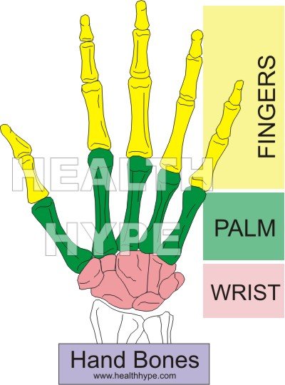
The human finger is mainly a bony structure with multiple joints giving it strength and flexibility. A digit includes the hand bones but these bones are not separated into individual appendages like a finger. Instead it is contained within a single structure – the hand. Tendons attached to muscles within the hand and forearm are responsible for the different movements of the fingers.
The five fingers include :
- 1st finger – thumb (polex)
- 2nd finger – index finger (digitus secundus manus)
- 3rd finger – middle finger (digitus medius)
- 4th finger – ring finger (digitus annularis)
- 5th finger – little finger / pinky (digitus minimus manus)
There are two surfaces of the fingers :
- Palmar surface (front of the hand) continuous with the palms of the hand.
- Dorsal surface (back of the hand) containing the fingernails at the tips.
Finger Bones
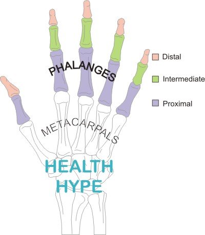
The finger bones are known as phalanges (singular ~ phalanx). There are 14 phalanges on each hand. All the fingers have 3 phalanges except the thumb which has 2 phalanges. Each phalanx in a finger is named according to its location :
- Proximal phalanx is the first finger bone lying next to the palm.
- Intermediate phalanx is the middle finger bone which is absent in the thumb.
- Distal phalanx is the last finger bone lying furthest away from the hand.
The hand bones are known as the metacarpals and correspond to the phalanges – the first metacarpal articulates with the proximal phalanx of the first finger.
Each phalanx has three parts – the base, shaft and head. The base of each phalanx articulates with the head of the preceding phalanx, except for the proximal phalanges (first finger bones) which articulate with the head of the metcaarpals (hand bones). The enlarged end of each phalanx (finger bone) being either the base or head is known as the knuckle bone.
Finger Joints
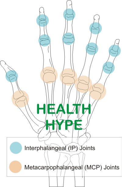
There are two types of finger joints, all of which are commonly referred to as knuckle joints :
- between the finger bones – interphalangeal joints (finger-finger joint)
- between the hand bones and first finger bones – metacarpophalangeal joints (hand-finger joint)
There are two interphalangeal joints (IP joints) on each finger, except for the thumb which has one.
- The first IP joint between the proximal and intermediate phalanges is known as the proximal interphalageal joint (PIP joint).
- The second IP joint between the intermediate and distal phalanges is known as the distal interphalangeal joint (DIP joint).
There is only one metacarpophalangeal joint (MCP joint) which lies between the proximal phalanx and metacarpal (hand bone). The ends of the bones involved in the joint is lined with articular cartilage. Synovial membranes line the joint and a tough capsule surrounds the joint.
Muscles and Movements
The muscles that control the movement of the fingers are located in the forearm and hand. Tendons running from these muscles attach to various points on the finger bones. When the muscle contracts, the tendon is pulled and the finger moves at the respective joint. Therefore these muscles, although not in the finger, should be discussed briefly.
There are two ways in which the muscles controlling the fingers can be classified. The first is by location.
- Intrinsic muscles which are located in the hand. There are three groups – thenar and hypothenar, interossei and lumbrical muscles.
- Extrinsic muscles which are located in the forearm. There are two groups – extensors and flexors.
The other classification of these muscles is by the movement of the fingers.
- Flexion of the fingers where the fingers move towards the palm. The muscle groups responsible are the thenar and hypothenar (intrinsic) and the flexors in the forearm (extrinsic).
- Extension of the fingers where the fingers straighten out by moving away from the palm. The muscle groups responsible are the interossei and lumbrical muscles (intrinsic) and the extensors in the forearm (extrinsic). Volar ligaments over the palmar side of the IP joints prevents hyperextension.
Other movements like abduction where the fingers fan out, away from the middle finger, and adduction where the fingers move in towards the middle finger, are also controlled by certain intrinsic and extrinsic muscles. Circumduction is when the finger moves in a circular manner.
Nerves of the Fingers
Nerves send signals from the brain to the muscles (motor nerves) causing it to contract or from receptors in the fingers to the brain (sensory nerves) to enable the different sensations. The motor nerves supplying the muscles controlling the fingers is not discussed here since these muscles are located in the hand and forearm. It involves the median, ulnar and radial nerves.
The skin of the fingers are supplied by different nerves as follows :
- Median nerve – palmar surface, tips and nail beds of the thumb, index, middle and half of the ring fingers.
- Ulnar nerve – palmar and dorsal (back of the hand) surface of the other half of the ring finger and the little finger.
- Radial nerve – dorsal surface (excluding the tips) of the thumb, index, middle and half of the ring fingers and web between thumb and index fingers.


