Hydronephrosis (Kidney Swelling with Urine Blockage)
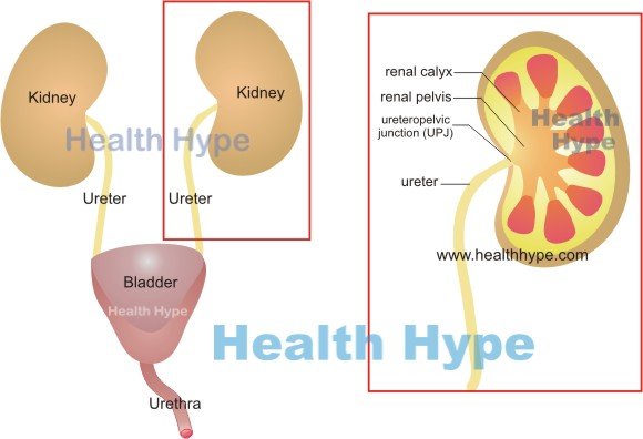
The kidneys are the two bean-shaped organs located on either side of the abdominal cavity. It is responsible for the expulsion of waste substances from the body and also has an effect on blood pressure and other vital functions. The human kidneys filter blood throughout the day and night thereby producing some 1 to 2 liters of urine daily on average. The urine is then passed down the ureters on either side and drains into the urinary bladder. Here it is stored until there is the need to expel this urine.
The human bladder can hold about 500ml of urine and once full, the body is signaled that this accumulated urine needs to be passed out into the environment. When the setting is appropriate, the sphincters open and the muscular wall of the bladder contracts tightly thereby propelling the urine into the urethra and out into the exterior. Any blockage either in the upper or lower urinary tract can hamper the passage of urine. If the urine produced then accumulates to a significant degree, it can cause expansion of the urinary tract above the site of the obstruction.
What is hydronephrosis?
Hydronephrosis is the distention of certain parts of the kidney due to the accumulation of urine arising from an obstruction to its outflow. Another related term – hydroureter – is distention of the ureter due to excessive amounts of urine collecting within in. Stasis of urine increases the risk of urinary tract infections or kidney stones. Severe or untreated cases of hydronephrosis can cause permanent kidney damage and lead to kidney failure. Hydronephrosis and hydroureter can occur in adults, children, newborn babies and even in the fetus often for different reasons. Most instances of hydronephrosis are unilateral meaning that it affects only one kidney although it can occur on both sides (bilateral).
Reasons for Hydronephrosis
The kidneys are constantly producing small amounts of urine by filtering blood. Water and different substances within the blood pass into the glomerulus of the nephron – the basic functional unit of the kidney. This glomerular fluid then begins its passage along the tubule of the nephron. Different substances are pushed into the tubule or extracted from the tubular fluid until the resulting fluid passes out into the renal calyx. This fluid is known as urine. The calyces (singular ~ calyx) converge to form the renal pelvis – a single collecting area of urine within the kidney. It then empties through into the ureter. This passage of urine is aided by rhythmic muscular contractions in the wall of these structures.
Should an obstruction arise, usually within the upper parts of the ureter, the passage of urine is hampered. The urine then collects above the site of the obstruction. Eventually the backward pressure of the urine causes the ureter, renal pelvis and calyx to stretch (distend). As the condition progresses the pressure buildup extends into the nephron thereby hampering normal glomerular filtration. This continues to have a knock on effect if left untreated and eventually damages the functional apparatus of the kidney. In severe cases the kidney tissue is irreversibly damaged.
However, the ureter and renal pelvis is able to distend to a significant degree before this damage to the kidney tissue arises. The swollen portions may become tortuous (twist) and can further complicate the existing obstruction.
Causes of Hydronephrosis
The causes of hydronephrosis can be classified as intrinsic (within the urinary tract) or extrinsic (cause lies outside of the urinary tract). Most causes lie within the upper parts of the ureter but hydronephrosis can rise with a blockage anywhere in the urinary tract even within the urethra.
- Kidney stones that lodge within the ureteropelvic junction (where the ureter joins the kidney) or in the ureter.
- Blood clot in the renal pelvis or ureter.
- Strictures (narrowing) of the ureter whether due to a problem within the ureter or around it. Scar tissue is a common cause of ureteral strictures.
- Tumors in the ureter (intrinsic cause) or in structures around the ureter that compress it (extrinsic cause).
- Pregnancy where the growing uterus presses against the ureter.
There are various other causes of a blocked ureter that may lead to hydronephrosis. Less commonly the obstruction lies within the lower parts of the urinary tract like the distal ureter, bladder and urethra.
- Cancer of the bladder.
- Cancer of the prostate (men) or cervix and uterus (women).
- Enlarged prostate (men) or rectum with fecal impaction.
- Bladder stones
- Neurogenic bladder
- Ureterocele
- Cystocele
- Urethral strictures (narrow urethra)
An obstruction in the bladder or urethra is more likely to cause bilateral hydronephrosis (both kidneys).
Pregnancy
Hydronephrosis of pregnancy occurs as a result of the growing uterus compressing the ureters. It can lead to bilateral hydronephrosis. The hormonal changes that occur with pregnancy also reduces the muscular contractions in the ureter that help with the movement or urine down into the bladder
Newborns
There are several reasons for hydronephrosis in newborn babies (pediatric hydronephrosis). The most common cause is ureteropelvic junction (UPJ) obstruction due to a developmental defect.
Fetus
Fetal hydronephrosis occurs in the unborn baby and may remain undetected until after birth. However, modern diagnostic techniques may be able to detect this problem in the fetus. It arises with different conditions like kinking of the ureter or reflux of urine from the bladder into the ureter (vesicoureteral reflux).
Signs and Symptoms
The signs and symptoms of hydronephrosis depends on the degree of obstruction, whether one or both kidneys are affected and the duration of the condition. It may be asymptomatic in the early stages. Signs and symptoms of hydronephrosis includes :
- Flank pain (upper flank) on the affected side correlating with position of the kidney. Read more on kidney pain location. The pain may be severe and constant or intermittent.
- Oliguria (reduced urine flow) or rarely anuria (complete absence of urine) if both ureters are affected (bilateral hydronephrosis). When it is one-sided (unilateral), the urine flow remains normal.
- Nausea and vomiting (sometimes).
- Abdominal pain (central or generalized).
- Leg swelling if there is bilateral hydronephrosis.
- Blood in the urine that arises with certain causes of hydronephrosis.
There may be other symptoms such as muscle spasms and irregular heart rhythm (arrhythmia) with chronic hydronephrosis which otherwise remains fairly asymptomatic until the condition becomes severe.
Diagnosis
Various diagnostic techniques may be employed when hydronephrosis is suspected or there are urinary symptoms of an unknown cause. This may include :
- Ultrasound
- Bladder catheterization
- Intravenous pyelography (IVP)
- CT (computed tomography) scan
- MRI (magnetic resonance imaging)
- Blood and urine tests
Treatment of Hydronephrosis
Therapeutic measures should be directed at the cause of the obstruction. Once this is resolved the urine flow will return to normal. Irreversible damage to the kidney usually happens after several weeks and only when both kidneys are affected (bilateral hydronephrosis). This allows for some leeway in treating the cause and awaiting the restoration of normal urine output.
The various causes need to be treated accordingly, for example kidney stones may need to be removed with procedures such as lithotripsy. Specific procedures may be necessary to restore urine flow when the cause is persisting, the symptoms are severe or there is risk of complications such as kidney failure. This mainly involves one of two procedures :
- Nephrostomy where a tube is inserted through the skin into the kidney for urine drainage.
- Ureteral stent where a tube is used to connect the kidney to the bladder thereby passing the ureter.
References
Hydronephrosis and hydroureter. Emedicine Medscape
Hydronephrosis. Merck Manuals


