Left-to-Right Cardiac Shunts (Heart) Types and Symptoms
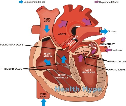
Congenital heart defects arise in the fetal stage in life when the development of the heart and/or great blood vessels is disrupted in some manner. This leads to a structural abnormality in the heart or vessels which depending on the type and extent may case mild to severe symptoms or can even be life threatening. The heart is a a muscular pump with four chambers – two atria that receive blood and two ventricles that push out blood. The atrium and ventricle on each side are separated by heart valves which open and close at different stages of the cardiac cycle. Similarly the ventricles and arteries communicating it are separated by valves.
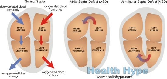
However, the atria and ventricles are kept separate from each other by the atrioventricular (AV) septum. This means that the blood from the right atrium or ventricle cannot mix with the blood in the left atrium or ventricle. The septum is essential to keep oxygen deficient blood in the right chambers of the heart separate from the oxygen rich blood in the left chambers. In certain types of congenital heart defects, the septum is compromised thereby allowing for a mixing of the blood.
What is a cardiac shunt?
A cardiac shunt, or heart shunt, is abnormal communication between chambers or blood vessels that allows for the passage of blood. Many of the more common types of congenital heart defects are due to malformations that lead to shunting of blood. This allows for blood on one side of the heart to enter the other side through an abnormal channel.
Cardiac shunts are categorized according to the direction of abnormal flow. In this manner, a shunt may either be a :
- Left-to-right shunt – oxygenated blood from the left side of the heart enters the chambers and conduits containing deoxygenated blood (pulmonary circulation).
- Right-to-left shunt – deoxygenated blood from the right side of the heart enters the chambers and conduits containing oxygenated blood (systemic circulation).
Left-to-right shunts are the most common type of congenital heart defect. A left-to-right shunt may not seem like a serious condition because oxygen-rich blood from the left is entering the areas containing oxygen-deficient blood on the right. This blood is then sent to the lungs for oxygenation. However, the danger with a left-to-right shunt is the structural changes in the heart and blood vessels that arises with the additional blood in the right side of the heart.
Types of Left-To-Right Heart Shunt
The pressure in the right side of the heart is lower and the heart walls and related vessels have developed to tolerate the pressure. With a left-to-right shunt, oxygenated blood from the left side of the heart passes to the right side. This increases the volume of blood in the pulmonary circulation and therefore raises the pressure. The walls and vessels thicken to accommodate this greater pressure and leads to similar structural changes as is seen with hypertension (high blood pressure). More importantly though, with time the structural changes may lead to a state where the flow is no longer from left-to-right but rather from right-to-left.
The four most common types of left-to-right cardiac shunts are :
- Atrial septal defect (ASD)
- Ventricular septal defect (VSD)
- Patent ductus arteriosus
- Atrioventricular septal defect
Atrial Septal Defect
An atrial septal defect (ASD) is an abnormal opening between the right and left atria. This fixed opening in the atrial septum is result of incomplete closure of the atrial septum and allows blood from the left atrium to pass into the right atrium. It may be of three types known as secundum atrial septal defects (the most common), primum anomalies and sinus venous defects. ASD’s tend to become symptomatic only in adulthood.
Ventricular Septal Defect
A ventricular septal defect (VSD) is an abnormal opening between the right and left atria. It is a fixed opening formed by incomplete closure of the ventricular septum. This allows from blood from the left ventricle to pass into the right ventricle. VSD’s are the most common type of congenital heart defect accounting for about 30% of these defects. It is often associated with other types of heart defects. Large openings may cause severe symptoms from birth while smaller openings may close spontaneously and only cause symptoms in adulthood if it remains open.
Patent Ductus Arteriosus
In fetal life, blood does not need to be oxygenated in the lungs because oxygenated blood is derived from the mother. However, the fetus still has a fully functional heart with separate chambers. To prevent blood from being sent to the lungs, two possible conduits exist. The first is a small channel known as a ductus arteriosus which shunts blood from the pulmonary artery (that leads to the lungs) to the aorta (which carries blood to the rest of the body). This shunt is normal in fetal life. However it may sometimes persist after birth.
A patent ductus arteriosus is the persistence of the channel between the pulmonary artery and aorta after birth. This allows oxygenated blood in the aorta to enter the deoxygenated blood in the pulmonary artery which is headed to the lungs. The severity of the condition depends on the size of the shunt but is usually asymptomatic in infants.
Atrioventricular Septal Defect
An atrioventricular septal defect (AVSD) is the incomplete closure of the septum between the left and right chambers of the heart. It may be partial or complete. AVSD is associated with atrioventricular (AV) valve defects – tricuspid valve of the right side or mitral valve of the left side. This condition is seen more frequently with Down’s syndrome.
Signs and Symptoms of Left-to-Right Shunts
Babies born with these heart defects may be asymptomatic for years or right up until adulthood. However, in severe cases the symptoms may be prominent from birth. Typically features like cyanosis (bluish discoloration of the skin) are not present with a left-to-right shunt but becomes prominent if a right-to-left shunt develops at a later stage.
- Heart murmurs
- Arrhythmias
- Enlargement of the right side of the heart
- Enlargement of the pulmonary artery
- Dizziness
- Fatigue
- Exertional dyspnea – shortness of breath with physical activity
- Learning difficulties in childhood


