Pancreas Location, Anatomy, Parts, Pancreatic Duct and Pictures
What is the pancreas?
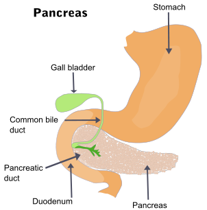
The pancreas is an elongated lobular gland extending from the C-loop of the duodenum to the hilum of the spleen. Due to its shape, the pancreas is divided into a head, neck, body and tail for descriptive purposes. An extensive ductule network and large pancreatic duct empties into the common bile duct.
The multi-function gland has both endocrine and exocrine functions. The endocrine component releases hormones directly into the blood stream and play a major role in metabolism and a lesser role in digestion. The exocrine part of the pancreas releases digestive enzymes into the duodenum thereby facilitating chemical digestion. The pancreatic hormones and enzymes are discussed further under Pancreas Function.
Picture from Wikimedia Commons
Pancreas Anatomy
The pancreas measures about 20 centimeters in length (about 8 inches) and weighs between 75 to 90 grams (heavier in men). The bulk of the pancreatic tissue is dedicated to its exocrine function, while only 1% to 2% of the pancreas is responsible for the endocrine component.
The pancreas has two main types of cells – the islet of Langerhans responsible for the endocrine function and the acinar cells that produce and secrete digestive enzymes (exocrine). The acinar cells are arranged in clusters to make up the pancreatic acini which drain the enzymes into introlobular ductules that eventually merge to form the main pancreatic duct.
Arterial Supply
- Superior pancreaticoduodenal artery – branch of gastroduodenal artery
- Inferior pancreaticoduodenal artery – branch of superior mesenteric artery
- Pancreatic branches of splenic artery
Venous Drainage
- Pancreatic veins which empty into the superior mesenteric and splenic parts of the hepatic portal vein and splenic vein
Lymphatic Drainage
- Pancreatic lymph vessels empty into the pancreaticospenic lymph nodes or pyloric lymph nodes
Nerve Supply
- Sympathetic and parasympathetic fibers from celiac plexus and superior mesenteric plexus (vagus and abdominopelvic splanchnic nerves)
Pancreas Parts
Picture from Wikimedia Commons
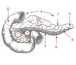
(1) head of pancreas, (2) uncinate process of pancreas, (3) pancreatic notch, (4) body of pancreas, (5) anterior surface of pancreas, (6) inferior surface of pancreas, (7) superior margin of pancreas, (8) anterior margin of pancreas, (9) inferior margin of pancreas, (10) omental tuber, (11) tail of pancreas, (12) duodenum
Head
The head of the pancreas is the enlarged part of the gland that is tucked against the the C-loop of the duodenum. It has an inferior projection known as the uncinate process. A groove on the posterosuperior surface (upper rear part) of the head accommodates the bile duct. The head of the pancrease rests against the inferior vena cava (IVC) that lies behind it (posteriorly). It makes contact with the right renal artery and left and right renal veins.
Neck
The neck of the pancreas is the short portion of the gland that connects the pancreatic head to the body. It measures about 1.5 to 2 centimeters (less than an inch) and lies adjacent to the pylorus of the stomach.
Body
The body of the pancreas is the continuation of the neck that begins to taper towards the tail. It makes contact with with the aorta, adrenal gland, left kidney and renal vessels.
Tail
The tail of the pancreas extends to the hilum of the spleen. The narrowness as well as distance away from the head mass makes it relatively mobile. It lies in front of the left kidney (anteriorly).
Pancreatic Duct
The main pancreatic duct receives pancreatic juices from the intralobular ductules. It starts at the tail of the pancreas and runs all the way to the head. Here it joins with the common bile duct at a dilate portion known as the ampulla of Vater (hepatopancreatic duct). This is the terminal end of the duct systems (pancreatic and bile) and the contents from these ducts are then emptied into the duodenum. It is not uncommon for the pancreatic duct to remain separate from the common bile duct and both may empty directly into the duodenum.
The pancreatic duct sphincter is a smooth muscle sphincter that controls the outflow of the pancreatic juice into the ampulla. In turn, the sphincter of Oddi (hepatopancreatic sphincter) controls the bile and pancreatic secretions into the duodenum.
The pancreas also has an additional duct for the outflow of pancreatic juices. This is known as the accessory pancreatic duct and most of the time it communicates with the main pancreatic duct. It can however occur independently and remain unconnected to and may even be larger than the main pancreatic duct.
Pancreas Location
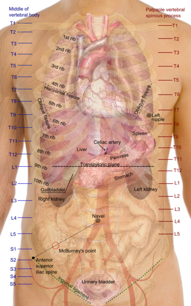
The pancreas is located in the upper part of the abdomen, primarily in the epigastrium and extending into the left hypogastrium. It lies retroperitoneally traversing the transpyloric line (refer to picture below) at the level of the L1 and L2 vertebrae. Abdominal organs and structures related to the pancreas includes :
- stomach which is situated in front of the pancreas (anterior),
- inferior vena cava (IVC) and abdominal aorta situated behind the pancreas (posterior).
Picture from Wikimedia Commons


