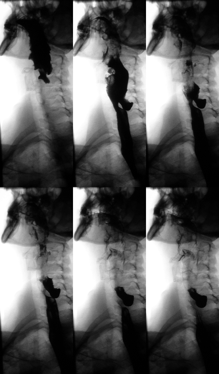
Zenker Diverticulum
What is Zenker diverticulum?
Zenker diverticulum is an outpouching of the wall of the upper esophagus (gullet). It is rare and almost only seen in the elderly. Outpouchings are protrusions of the wall of a hollow cavity. It can occur in any part of the gut and a more common type is seen in the large intestine. Zenker diverticula (singular ~ diverticulum) need to be repaired surgically. When left untreated it can lead to complications, some of which are severe like pneumonia. A Zenker diverticulum or Zenker’s diverticulum is named after by Professor Frederich Albert von Zenker who described this type of diverticulum in 1877.
Zenker Diverticulum Location
Anatomy of the Esophagus
The esophagus is a long hollow tube that runs from the pharynx (throat) to the stomach. It has a thick wall with strong muscles that push food down to the stomach. The esophagus runs from the neck, down through the chest cavity and into the abdomen. There are two sphincters in the esophagus :
- Upper esophageal sphincter (UES) which allows food to pass from the throat into the esophagus.
- Lower esophageal sphincter (LES) which controls the movement of food into the stomach.
Both these sphincters are under involuntary control. It opens when food is approaching the sphincter and prevents backward movement of food and fluids.
Protrusions of the Esophagus
Protrusions can occur in the wall of any hollow cavity and not only the esophagus. When it occurs in a blood vessel then it is known as an aneurysm. If it occurs in the alimentary tract then it is known as a diverticulum or hernia. The more common diverticula are seen in the lower gut, especially the colon and last part of the small intestine (ileum). Esophageal diverticula can occur in the upper, middle and lower gut where it is known as :
- Zenker diverticulum – just above the UES.
- Traction diverticulum – around the middle of the esophagus.
- Epiphrenic diverticulum – just above the LES.
Diverticula are essentially evaginations where tissue bulges outwards, as opposed to invaginations where tissue protrudes inwards. There are three ways to classify these protrusions :
- True diverticula contains all layers of the intestinal wall, from the most superficial to the deepest.
- False diverticula where only the inner lining and the layer just beneath it are involved.
- Intramural diverticula where an inner layer within the wall, like the submucosa, is only involved.
Position of Zenker Diverticulum
Diverticula can occur anywhere along the length of the esophagus. A Zenker diverticulum is located just above the upper esophageal sphincter and technically involves the pharynx (throat). Therefore it is also known as a pharyngeal or pharyngoesophageal diverticula. A Zenker diverticulum forms on the back (posterior) wall of the pharynx just above the UES. It is considered to be a “false” diverticulum because only the inner lining of the wall (mucosa) and the tissue layer just underneath it (submucosa) is involved.
Zenker Diverticulum Pathphysiology
Normal swallowing
It is important to understand the process of swallowing when considering how a Zenker diverticulum may arise. There are three phases of swallowing, one of which is voluntary while the other two are involuntary.
- Oral stage of swallowing where food is pushed to the back of the mouth is voluntary.
- Pharyngeal stage where the food is taken from the back of the mouth and pushed down the throat is involuntary.
- Esophageal stage where food is propelled all the way down to the stomach is involuntary.
Once voluntary swallowing is initiated and food moves from the back of the mouth to the throat, the rest of the process occurs “automatically” and cannot be stopped.
Weakening in the wall
In order for food in the throat to enter the esophagus, the upper esophageal sphincter (UES) must relax. This sphincter is made up by several muscles that surround it. The main muscle is the cricopharyngeus muscle. It is believed that a Zenker diverticulum arises when the relaxation of the cricopharyngeus muscle is disordered. This applies pressure on the esophagus and does not allow food in the pharynx (throat) from entering the esophagus properly.
Pressure builds up within the pharynx during swallowing as food is being pushed. However,the abnormal activity of the cricopharyngeus muscle causes the pressure within the pharynx to rise to a much higher level during swallowing. This causes a bulging of the wall to occur over time. Since the back wall (posterior wall) is the weakest portion, the outpouching develops here. It more specifically occurs in an area known as Killian’s triangle which is not supported by the muscles that normally surround the esophagus from the outside to form the upper esophageal sphincter (UES).
Zenker Diverticulum Causes
Although the possible mechanism by which a Zenker diverticulum may form is described above in a rather simplistic manner, the exact cause of this condition is unclear. For now it can be said that a Zenker diverticulum is formed by incoordination of the cricopharyngeus muscle which essentially fails to relax sufficiently during the swallowing process. However, there may be other factors that contribute to the condition. It is possible that there is some other cause of weakening of the wall which makes it prone to herniation.
Zenker Diverticulum Symptoms
The symptoms of a Zenker diverticulum depends on its size. Initially it may have little to no symptoms while it is small. Occasionally patients ignore the symptoms until it gets severe or leads to complications such as pneumonia.
Swallowing symptoms
These are the most common and obvious symptoms. It includes :
- Difficulty swallowing, specifically throat swallowing.
- Regurgitation of chewed but undigested food. Vomiting does not occur.
- Noisy swallowing.
Breath symptoms
Bad breath (halitosis) may be reported as food that is trapped in the diverticulum decomposes.
Voice symptoms
Vocal changes may be noticed in patients an is also dependent on aspiration, duration of time that the diverticulum existed and its size.
Other symptoms
Sometimes a Zenker diverticulum can cause a palpable mass in the neck. This is rare however, even with a large diverticulum. The presence of other symptoms are more likely to be related to the complications that arises with a Zenker diverticulum.
- Coughing and choking with aspiration.
- Chest pain, coughing and difficulty breathing with pneumonia.
Zenker Diverticulum Diagnosis
The report of symptoms by the patient should raise the suspicion of a Zenker diverticulum or sometimes other esophageal conditions like achalasia. The condition can be conclusively diagnosed with a barium swallow. A special substance known as barium sulfate is consumed and can then be tracked by x-ray as it passed down the throat and esophagus as is seen in the image. This test will conclusively confirm or exclude a Zenker diverticulum.
Zenker Diverticulum Treatment
A Zenker diverticulum cannot be treated with medication. It requires surgery to repair the outpouching. Although small pouches may not be repaired immediately, it should be monitored and surgery considered once symptoms develop or there are complications. There are two procedures that need to be conducted in repair surgery :
- Division of the cricopharyngeus muscle to relieve pressure. This procedure is known as a myotomy.
- Removal of the pouch (divertculum). This is known as a diverticulotomy.
Repair Surgery
A myotomy alone used to be done in the past but low relief of symptoms and the condition often recurred. There are three types of surgical procedures to remove or seal the pouch and divide the muscle.
- Diverticulectomy with cricopharyngeal myotomy
- Diverticulopexy with cricopharyngeal myotomy
- Endoscopic myotomy
References

