Tongue | Anatomy, Parts, Pictures, Diagram of Human Tongue
The human tongue is a muscular organ that is covered by a thin mucous membrane. It lies partly in the mouth cavity and partly in the oropharynx. It is highly mobile and can be shifted into a number of different positions and also assume various shapes. The tongue’s primary function is often seen as that of being the organ of taste, however, its role in various other activities is also crucial.
Functions of the Tongue
- Taste. The taste buds, the sensory receptors for taste, are located on the tongue.
- Speech. The movements of the tongue are crucial for articulation.
- Chewing and swallowing. The tongue helps the teeth and other parts of the mouth with chewing food and passing it down the throat as the first part of the swallowing process.
- Cleaning. The movements of the tongue dislodge food particles stuck between the teeth, gum and cheek so that it can be spat out or swallowed.
Parts of the Tongue
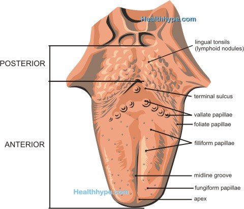
The top of the tongue (superior surface) has a V-shaped line known as the terminal sulcus that divides the tongue into the anterior and posterior surfaces.
- The anterior surface is made up of the apex at the tip and body.
- The posterior surface is made up entirely of the root.
The inferior surface of the tongue (underside) is also made up of the body and apex.
Picture : Side View of the Human Tongue
Root
- Located between the hyoid bone and mandible.
- Dorsal portion sits in the oropharynx.
- Attaches the tongue to roof of the mouth.
Body
- Makes up the anterior two-thirds of the tongue.
- Rough surface due to the lingual papillae.
- Surrounded by anterior and lateral teeth.
- Mobile portion of the tongue.
Apex
- Also known as the tip, is the anterior one-third of the anterior tongue surface.
- Rests against the incisor teeth.
- Highly mobile.
Surface Features of the Tongue
The lingual papillae contain the taste buds and are located on the anterior surface (body and tip) of the tongue :
- Vallate papillae are large and flat papillae arranged in a V-shaped row just in front (anterior) of the terminal sulcus.
- Foliate papillae are poorly developed folds on the side of the tongue.
- Filiform papillae are long, conical, pinkish gray projections that are sensitive to touch.
- Fungiform papillae are pink to red spots distributed between the filiform papillae and are most dense at the apex and margins of the tongue.
The posterior surface of the tongue has no lingual papillae but has a rough surface due to the presence of lymphoid nodules.
The midline groove divides the anterior part of the tongue into the left and right parts.
The inferior surface is connected to the floor of the mouth by a fold known as the frenulum. A visible vein on either side of the frenulum is known as the deep lingual vein.
The sublingual papillae (caruncle) is located on either side of the base of the frenulum and it is the opening for the ducts of the submandibular gland (salivary glands).
Muscles of the Tongue
The tongue is a muscular mass and although it is made up of several muscles, all act in conjunction with each other to perform various movements. The tongue muscles can be divided into the intrinsic and extrinsic groups. Broadly the intrinsic muscles can alter the shape of the tongue while the extrinsic muscles change the position of the tongue.
Intrinsic Muscles
These four muscles originate and terminate in the tongue and do not attach to any bone.
- superior longitudinal
- inferior longitudinal
- transverse
- vertical
The superior and inferior longitudinal muscles can retract the tongue thereby making it short and thick. The transverse and vertical muscles protrude the tongue out of the mouth thereby making it long and narrow.
Extrinsic Muscles
These four muscles originate outside the tongue where it is attached to bone.
- genioglossus
- hyoglossus
- styloglossus
- palatoglossus
These muscles are primarily responsible for moving the tongue but also play a role in altering the shape of the tongue.
Nerve Supply to the Tongue
Motor
All muscles are innervated by hypoglossal nerve (cranial nerve 12, CN XII) except the palatoglossus muscle which is supplied by the pharyngeal plexus of CN X.
Sensory
Taste
- Anterior Two-Third (excluding vallate papillae) : chorda tympani (branch of CN VII)
- Posterior One-Third (including vallate papillae) : lingual branch of glossopharyngeal nerve (CN IX)
Touch and Temperature
- Anterior Two-Third : lingual nerve (branch of CN V)
- Posterior One-Third : lingual branch of glossopharyngeal nerve (CN IX)
Blood Supply to the Tongue
Oxygenated blood to the tongue is carried via the lingual artery which arises from the external carotid artery. The body of the tongue is supplied by the deep lingual arteries, while the root of the tongue is supplied by the dorsal lingual arteries.
Dexoygenated blood is drained out of the tongue by the lingual veins – dorsal and deep. The deep lingual vein which starts at the apex of the tongue is visible on the inferior side of the tongue on either side of the lingual frenulum. The deep lingual vein may drain into the sublingual vein. The dorsal and sublingual veins in turn empty into inferior jugular vein. Sometimes the dorsal and deep lingual veins may join to form a common lingual vein.
Lymphatic Drainage of the Tongue
- Root – superior deep cervical lymph nodes
- Body (medial part) – inferior deep cervical lymph nodes
- Body (lateral parts) – submandibular lymph nodes
- Apex, frenulum – submental lymph nodes


