Bladder (Urinary), Anatomy, Location, Parts and Pictures
The urinary bladder is a hollow muscular organ located in the lesser pelvis when empty. It serves as a reservoir for urine and can stretch considerably to store close to a maximum of 500 milliliters of urine. The average full bladder that is not overly distended contains about 350 milliliters of urine. It receives urine produced in the kidneys via the ureters and passes it out into the external environment through the urethra.
Anatomy of the Urinary Bladder
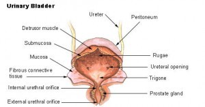
The empty urinary bladder is somewhat tetrahedral in shape – like a three sided pyramid with a triangular base (illustrated in diagram). This gives the bladder one superior surface (top), two inferolateral surfaces (sides) and a posterior surface (back). The external aspect of the superior surface of the bladder is covered by peritoneum.
The internal surface of the bladder is lined with mucosa, which is folded to form rugae. This makes the internal surface of the bladder rough except for the smooth area of the trigone. The bulk of the bladder wall is composed of the detrusor muscle. which is smooth muscle and therefore under involuntary control.
Parts of the Urinary Bladder
The apex of the bladder points forward towards the pubic symphysis, while the base (fundus) lies posteriorly, against the rectum in males or vagina in females. The neck of the bladder is the inferior aspect where the bladder walls narrow and converge towards the urethra like a funnel. This directs urine into the urethra.
The body of the bladder is the largest part lying between the apex, fundus and neck of the bladder. The trigone of the bladder is a triangle region on the posterior wall. The two ureteric orifices (ureteral opening where the ureters enter) and internal urethral orifice (where the urethra begins) mark the three points of the trigone. It has a slight elevation known as the uvula of the bladder.
The parts of the detrusor muscle towards the neck of the bladder form the internal urethral sphincter. Some fibers run around the neck (radially) for better bladder control. This sphincter closes during ejaculation to prevent backward flow of semen into the bladder in men. The internal urethral sphincter is under involuntary control. The external urethral sphincter which is under voluntary control (skeletal muscle) is formed by the urogenital diaphragm.
Picture from Wikimedia Commons
Blood Supply to the Bladder
- Arteries
- Mainly superior vesical arteries and to a lesser extent, the obturator and inferior gluteal arteries
- Males – inferior vesical arteries
- Females – vaginal arteries
- Veins
- Vesical venous plexus >> internal iliac veins or into the internal vertebral venous plexus
- Males – vesical plexus communicates with the prostatic plexus
- Females – vesical plexus communicates with the vaginal/uterovaginal plexus
Nerve Supply to the Bladder
- Sympathetic – from upper lumbar spinal cord and inferior thoracic nerves through the hypogastric plexus
- Parasympathetic – from sacral spinal cord through the pelvic splanchnic nerves and inferior hypogastric plexus
Location of the Urinary Bladder
As it fills with urine, the bladder rises up into the greater pelvis. In infants and children, the bladder is contained within the lower abdomen when empty or full and drops into the greater pelvis around the age of 6 years. After puberty it descends into the lesser pelvis.
Picture from Wikimedia Commons
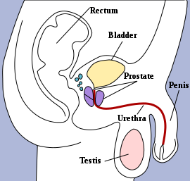
The bladder lies below the level of the umbilicus although in some individuals it can rise to this level when full. Its location is best understood in relation to surrounding organs and structures :
- MEN
- Superior (above) – peritoneal cavity
- Posterior (back) – rectum (separated by the rectovesical septum)
- Inferior (below) – prostate gland
- Anterior (front) – linea alba and pubic symphysis (separated by the retropubic space)
- WOMEN
- Superior (above) – peritoneal cavity and uterus (separated by the vesicouterine pouch)
- Posterior (back) – vagina
- Inferior (below) – perineal membrane
- Anterior (front) – linea alba and pubic symphysis (separated by the retropubic space)


