Blood Vessels (Artery, Vein) Structure, Function, Inflammation
What are blood vessels?
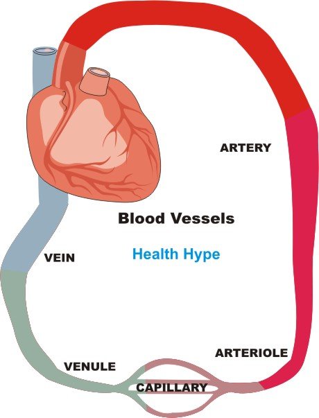
Blood vessels are the specially designed tubes that carry blood throughout the body. The vessels that carry oxygen-rich blood from the heart to the different areas of the body are known as arteries (singular ~ artery) while those vessels that carry oxygen-deficient blood back to the heart are known as veins (singular ~ vein).
The size of the blood vessels, both arteries and veins, differ throughout in the body and can be broadly categorized as :
- Artery – large (elastic) arteries, medium (muscular) arteries, small arteries and arterioles
- Vein – large veins, medium-sized veins, small veins and venules
The capillaries are the smallest segments of the circulatory system and are about as wide as the diameter of a red blood cell. It is the area of the gas exchange between the blood and tissue spaces.
Structure of the Blood Vessels
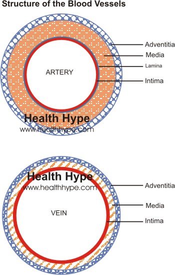
The structure of the blood vessels are very similar although variations may be seen due to functional differences of arteries and veins. The walls of the arteries are thicker and more muscular and elastic than the veins. This allows it to control the blood flow to different areas of the body and withstand the high pressure of blood leaving the heart.
The three layers of blood vessel walls include :
- Intima (tunica intima – inner)
- Media (tunica media – middle)
- Adventitia (tunica adventitia – outer)
Diagram
Both the arteries and veins have these three layers – it is more prominent in the arteries, especially the large and medium arteries and less defined in the veins. The tunica medica, which is the very muscular middle layer in arteries, is thinner and less muscular in veins. The veins also lack the elastic internal lamina that lies between the intima and media in the arteries. The tunica adventitia of the arteries are much thicker than that of the veins so that the artery can withstand the high pressure blood. The artery walls can be so thick that it may need its own blood supply known as the vaso vasorum.
The capillaries however are quite simple in structure. These vessels are very thin to enable gas exchange – it is usually just a single endothelial cell layer in thickness (similar to the intima), lacks a media layer and there may be some supporting connective tissue.
Functions of the Blood Vessels
- Arteries carry oxygenated and nutrient-rich blood to the body’s tissues from the heart. Dilatation and constriction of the arteries can alter blood pressure and cardiac output.
- Veins carry deoxygenated blood and wastes from the tissues to the liver and heart. The lympathic vessels also empty into the veins thereby returning any excess fluid in the tissue spaces back into the circulation.
- Capillaries allow for gas, nutrient and waste exchange between the blood and tissue spaces.
Blood Vessel Inflammation
Vasculitis is a broad term for inflammation of the blood vessel wall. There are some 20 types of vasculitis. It may occur in a vessel of any size although certain types of vasculitis tend to affect arteries of a specific size. Some forms of vasculitis also have a tendency to affect very specific blood vessels. Vasculitis is also known as arteritis or angitis and the term vasculiltides is used to describe the many types of vasculitis.
How does vasculitis occur?
Vasculitis may be due to infectious or non-infectious causes. In infectious vasculitis, pathogenic microorganisms infiltrate the blood vessel wall, although certain systemic infections may trigger inflammation in the vessel wall without directly infiltrating the site. Non-infectious vasculitis may be due to a number of causes from mechanical to chemical factors, drugs and irradiation. However, immune-mediated vasculitis appears to be one of the most common causes of non-infectious vasculitis.
Immune-Mediated Vasculitis
There are three mechanisms that result in immune-mediated vasculitis.
- Immune complex-associated vasculitis
- Antineutrophil cytoplasmic antibodies
- Anti-endothelial cell antibodies
While the mechanisms may differ, all these types of immune-mediated vasculitis are related to a hypersensitivity reaction. The triggers may vary from drugs to infectious agents or genetic disorders and in some cases of immune-mediated vasculitis, the cause is unknown.
Inflammation may arise when antibodies target certain layers of the vessel wall or when activated immune complexes deposit in the vessel wall and initiate the inflammatory process. This is often a systemic condition and other effects of immune-mediated hypersensitivity may also be present apart from the inflammation of the blood vessel wall.
Infectious Vasculitis
This is a result of an infection of the blood vessel wall. Pathogenic microorganisms, particularly bacteria or fungi, invade the vessel wall and this results in inflammation. The infection may spread from a neighboring site, occur as disseminated microorganisms travel through the bloodstream like in septicemia or when microorganisms are carried by a septic embolism.
Other infectious agents may be responsible for triggering immune-mediated vasculitis yet these microorganisms do not directly infiltrate the blood vessel wall.
Vasculitis Associated with Other Disorders
At time vasculitis may arise secondary to other disorders, particularly autoimmune diseases. This includes vasculitis associated with rheumatoid arthritis, SLE (systemic lupus erythematosus), inflammatory bowel disease, sarcoidosis, dermatomyositis, and cancer.


