Diaphragm (Human Thorax) Location, Anatomy, Function and Position
The Human Diaphragm
The word ‘diaphragm’ is used to describe several structures in the human body and in particular the thoracic, urogenital and pelvic diaphragms. The most important and largest of these structures is the thoracic diaphragm. It is a flat sheet of muscle that is responsible for breathing. The diaphragm essentially pulls and pushes against the lung causing it to expand with inhalation and contract with exhalation. The thoracic diaphragm also separates the organs in the thoracic cavity (chest) and abdominal (belly). By separating these cavities, the important organs like the lungs and heart can function properly in its own compartments. Some organs like the large blood vessels and esophagus (food pipe) pass across the diaphragm between the chest and belly through different openings. In this way the chest and abdomen can communicate with each other only at certain points.
Anatomy of the Diaphragm
The thoracic diaphragm is essentially a large muscle. Like all muscles, it has tendons that attach to the muscles and secure it. The diaphragm is attached to the inside of the ribs, to the back of the breastbone and the spine. The muscle fibers then meet in the middle of the diaphragm at the central tendon. Unlike other muscle tendons, the central tendon does not attach to any bone.
- Arteries that supply oxygen-rich blood to the diaphragm includes the phrenic arteries from aorta and internal thoracic arteries.
- Veins drain oxygen-deficient blood from the diaphragm through the phrenic arteries that drain into the suprarenal veins and inferior vena cava.
- Lymph is emptied from the diaphragm to the diaphragmatic lymph nodes and superior lumbar lymph nodes.
- Nerves that control the muscle action of the diaphragm are the phrenic nerves (C3 to C5) and sensation is through the phrenic, intercostal (T5 to T11) and subcostal (T12) nerves.

Picture from Wikimedia Commons
Shape of the diaphragm
The diaphragm is dome-shaped. The middle part of this dome is depressed just where the heart and its compartment, the mediastinum, are located. This makes the diaphragm a double dome, one on either side. It is the middle of these two side domes that move during respiration (breathing). During inhalation, the muscles contract and the diaphragm becomes almost flat. When the muscles of the diaphragm relax again it rises upwards to once again form the flat-topped dome shape.
Parts of the diaphragm
- Top of the diaphragm (superior surface) is convex and protrudes into the thoracic cavity.
- Bottom of the diaphragm (inferior surface) is concave and faces the abdominal cavity.
- Front of the diaphragm (middle part) is known as the sternal part. It attaches to the back of the breastbone (xiphoid process of the sternum).
- Back of the diaphragm (middle part) is the lumbar part. It communicates with the first three lumbar vertebrae.
- Sides of the diaphragm are the remaining parts, excluding the sternal and costal parts, which attach to the back of the last six ribs and its costal cartilages.
There are two bands known as crura (singular ~ crus) that arise from the lumbar vertebrae. There is the right crus and left crus. It runs through to continue into the central tendon and its surrounding muscle fibers. The right and left crus twist along its course to create two openings – one for the aorta and the other for the esophagus.
Openings in the diaphragm
There are several openings in the diaphragm also known as an aperture or hiatus, in the diaphragm. This allows for different structures and organs to run between the thoracic and abdominal cavity since the diaphragm separates these two cavities. There are three large openings and several smaller ones. From front to back is the caval opening, esophageal hiatus and aortic hiatus.
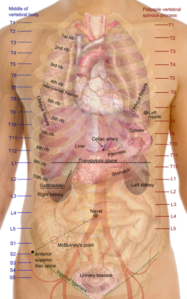
- Caval opening lies at the level of T8 and T9 vertebrae, slightly to the right of the midline of the diaphragm. The inferior vena cava runs through it, carrying oxygen-deficient blood to the heart. The right phrenic nerve and lymph vessels to the liver also pass through it.
- Esophageal hiatus is approximately at the level of the T10 vertebra. The esophagus (food pipe) runs through it allowing food from the throat to reach the stomach. The vagal trunks and a few blood and lymphatic vessels also pass through this opening.
- Aortic hiatus lies approximately at the level of the T12 vertebra. The aorta runs through it carrying oxygen-rich blood to the lower parts of the body. After passing through this opening, the aorta which was known as the thoracic aorta is now referred to as the abdominal aorta.

Picture from Wikimedia Commons
Functions of the Diaphragm
Breathing
The main function of the diaphragm is as a muscle of respiration, meaning that it aids with breathing. It is aided by other muscles and accessory muscles of breathing when a person experiences shortness of breath. When the diaphragm contracts, the lungs expand and air is inhaled. This occurs because it creates negative pressure within the pleural cavity around the lungs and the elasticity of the lungs allows it to expand thereby drawing in air. When the diaphragm relaxes, it restores the pressure in the pleural cavity and the lungs recoil thereby pushing out air (exhalation). Sudden and repeat contractions of the diaphragm, which are involuntary, leads to hiccups.
Compartmentalization
The diaphragm also separates the thoracic and abdominal cavities. This allows the heart and lungs to function in its own environment where it can maintain the relative pressure needed for its activity. It also prevents tightly packed abdominal cavity organs from making contact with the important thoracic structures – heart and lungs.
Blood Flow
When the diaphragm contracts, it pushes down the organs in the abdominal cavity. This action squeezes blood in the inferior vena cava thereby pushing the blood up towards the heart. It is further assisted by the decrease in the pressure within the thoracic cavity as the diaphragm flattens.
Diaphragm Location
The diaphragm is located at the junction of the thoracic and abdominal cavities about halfway down the chest behind the breasts. It has several organs lying immediately above and below it, with a few running through its openings and some even piercing it.

Picture from Wikimedia Commons
Position of the diaphragm
The position of the upper parts (dome) of the diaphragm changes with breathing. The periphery of the diaphragm is firmly attached to the breastbone, ribs and vertebrae and its position does not change.
- Periphery of the diaphragm originates at the level of the 11th and 12th ribs.
- Center of the diaphragm reaches as high as the 5th rib during expiration.
It is important to remember that the center of the diaphragm is pushed downwards when exhaling. It does not flatten entirely so it does not reach as low as the periphery. Fluid, a mass or other causes of raised pressure in the thoracic or abdominal cavity can push the middle of the diaphragm higher or lower. The liver sitting just under the right diaphragm causes it to be slightly higher than the left diaphragm. Read more on the .
Organs around the diaphragm
The main organs and structures around the diaphragm are as follows :
- Above the diaphragm lies the two lungs on either side and the mediastinum housing the heart at the middle.
- Below the diaphragm is the liver (right), pancreas (around the middle extending to the right), stomach (more towards the left), spleen (left) and kidneys (right and left sides).
- Front and around the diaphragm lies the breastbone and ribs.
- Back of the diaphragm lies the vertebral column.
Latest Updated on June 17, 2016 by admin
Published on December 17, 2011 by admin


