Duodenum Anatomy, Location, Parts and Pictures
What is the duodenum?
The duodenum is the first of the three parts of the small intestine and continues from the pylorus of the stomach. It is the shortest part of the small intestine, measuring approximately 25 centimeters in length and is also the most important site of digestion as the pancreatic enzymes and bile empty into the duodenum.
Anatomy of the Duodenum
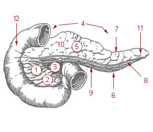
The duodenum runs a C-shaped course cupping the head of the pancreas and terminates at the duodenojejunal junction (flexure) where the jejunum (second part of the small intestine) arises. It can be divided into 4 parts :
- superior part – approximately 5 centimeters long
- descending part – 7 to 10 centimeters long
- inferior part – 6 to 8 centimeters long
- ascending part – approximately 5 centimeters long
Picture from Wikimedia Commons – (1 to 11) parts of the pancreas; (12) duodenum
Most of the duodenum is fixed in its position unlike the other parts of the small intestine that are fairly mobile. The duodenal papilla which is the opening of the hepatpancreatic ducts (bile + pancreatic ducts) is located in the descending part of the duodenum.
Blood Supply to the Duodenum
Arterial blood supply is via the celiac trunk and superior mesenteric artery. The gastroduodenal artery arising from the celiac trunk and its superior pancreaticoduodenal branch supply the superior part of the duodenum and portion of the descending part proximal to the duodenal papilla. The inferior pancreaticoduodenal artery arising from the superior mesenteric artery supplies the portion of the duodenum distal to the duodenal papilla.
Venous drainage is via veins that correspond to the arteries and empty directly into the hepatic portal vein or indirectly via the splenic and superior mesenteric veins.
Nerve Supply to the Duodenum
Innervation of the duodenum is via the celiac and superior mesenteric plexuses with nerves derived from the vagus and greater and lesser splanchnic nerves.
Lymphatic Drainage of the Duodenum
Anterior lymphatic vessels drain into the pancreaticoduodenal and pyloric lymph nodes, while the posterior lymphatic vessels drain into the superior mesenteric lymph nodes. Further drainage is into the celiac lymph nodes.
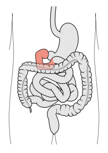
Picture from Wikimedia Commons
Location of the Duodenum
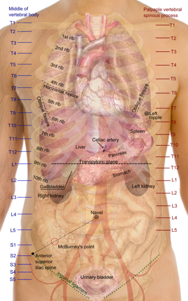
The duodenum lies in the upper abdomen, mainly in the epigastric region and extending into the umbilical quadrant of the abdomen . It starts at around the level of the L1 vertebra (superior part), runs downwards to the right of the L1 to L3 vertebrae (descending part), crosses the body of the L3 vertebra (inferior part) and courses upwards on the left of L3 and L2 vertebrae. Here it terminates at the duodenojejunal flexure which is about 2 to 3 centimeters to the left of the L2 vertebra.
Picture from Wikimedia Commons
The organs and structures surrounding the duodenum includes :
- Superior (above) – liver and gallbladder
- Anterior (in front) – gallbladder and transverse colon
- Posterior (behind) – inferior vena cava, right psoas major muscle and aorta
- Lateral – pancreas


