Myocardial Rupture (Heart Muscle Tear)
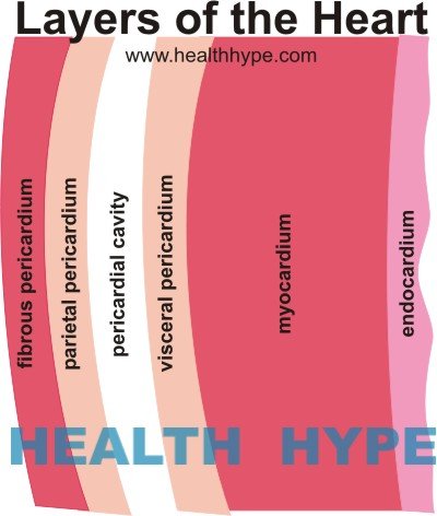
The heart wall has three layers and the thickest of these is the middle muscular layer. This is known as the myocardium. It is also the most active part of the wall since the heart is a pump that contracts and relaxes to circulate blood. Even a minor injury or damage of the heart muscle can affect blood circulation, lead to serious complications or rapidly progress to death.
What is myocardial rupture?
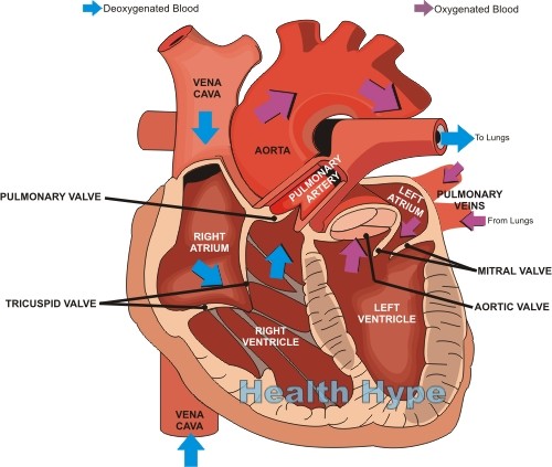
Myocardial rupture is a tear that occurs in the muscle layer of the heart wall. The tear can occur in the inner walls which divides the heart into separate chambers or on the outer wall which keeps the circulating blood within the heart. A myocardial rupture can also involve the tiny muscles that pull on the heart valves, the cords connected to these muscles and the heart valves, or even the flaps of the heart valves itself. The outlook for a myocardial rupture depends on where the tear occurs. Sometimes death is almost instant despite prompt medical attention. At other times medical treatment can prove to be life saving. However, the overall outlook for a myocardial rupture is poor.
Reasons for Tear in the Heart
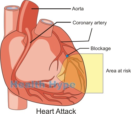
The strength of the heart wall is due to the muscle thickness and its ability to stretch and push blood out forcefully when the heart is filled. A rupture is more likely to arise when this wall is weakened or unable stretch like with death of the heart muscle. The accumulation of blood in the heart as its pumping ability is reduced applies force from the inside out. These several reasons are why a rupture is most like to occur a short while after a myocardial infarction (heart attack).
Here the blood supply to the heart wall is interrupted severely enough to allow a portion of the heart muscle to die. It is more likely to occur at the following sites (in order of decreasing frequency) :
- Outer wall of left ventricle (free wall)
- Outer wall of right ventricle (free wall)
- Inner wall between the ventricles (ventricular septum)
- Papillary muscles in the left ventricle
Sometimes more than one of these areas may tear and this is known as a double rupture. Although any mechanism that tears the heart muscle can also tear the inner lining (endocardium) and outer heart lining (epicardium), it is possible that the epicardium does not tear. In this case it contains the rupture but this cannot remain indefinitely as the epicardium does not have the same strength as the heart muscle. The outer sac covering the heart known as the pericardium can also contain the rupture in the same way as the epicardium. This is known as a pseudoaneurysm. It is a temporary seal and will not hold indefinitely.
Types of Myocardial Rupture
- Type I is a sudden slit-like tear that occurs a short while after a heart attack (myocardial infarction) usually within 24 hours.
- Type II is a slowly forming tear as the the damaged and dead heart muscle gradually erodes days after a heart attack, usually more than 24 hours afterwards.
- Type III is where the portion of the heart wall balloons (aneurysm) and eventually this aneurysm bursts.
Myocardial Rupture Causes
Heart Attack
A heart attack (acute myocardial infarction) is the most common cause of a myocardial rupture. The dead heart muscle is prone to tearing. A rupture may occur in as many as 10% of all heart attack cases. It is more common among females and in patients over the age of 60 years.
Heart Injury
There are many different mechanisms by which the heart may be injured.
- Blunt trauma where the heart may get compressed with a severe blow to the chest like during a car accident.
- Penetrating injury like in a gunshot or stab wound where the wound penetrates the chest wall, outer sac (pericardium) and then heart.
- Iatrogenic where the injury occurs during diagnostic investigation or surgical procedures.
Heart Infection
- Infective endocarditis
- Myocardial abscess rupture
- Acute myocarditis
- Echinococcal cysts
- Tuberculosis
Heart Tumors
- Primary tumors where the tumor arises from the heart tissue. It is more likely in malignant tumors (cancers) than benign tumors (non-cancerous).
- Secondary cancers where the cancer cells from a malignancy elsewhere in the body spreads to the heart (metastatic cancer).
- Lymphoma
- Acute myeloblastic leukemia
Other causes
- Aortic dissection
- Sarcoidosis
Heart Muscle Tear Symptoms
The symptoms can vary depending on where the tear occurs. A tear in the inner walls can cause mixing of blood between the heart chambers. A tear in the outer wall can cause blood to accumulate around the heart and prevent it from expanding. Rupture of the papillary muscles allows for backward flow of blood during heart contraction. Symptoms may not always develop immediately or be obviously detectable, even in a small but significant number of cases that arise with heart injury.
Some of the symptoms that may occur includes :
- Chest pain
- Rapid or slow heart rate
- Distended neck veins
- Muffled heart sounds, murmurs and other abnormalities of heart sounds
- Arrhythmia – irregular heart rate
- Pulmonary edema – fluid accumulation in the lung
- Signs of shock
Myocardial Rupture Diagnosis
There are various tests that may be conducted for a myocardial rupture. Some of these tests on its own will not conclusively indicate a tear unless confirmed with other investigations. Heart attack patients should be closely monitored for a myocardial rupture. It should also be suspected following a gunshot, stab wound or blunt injury to the chest. The presence of the symptoms above and the patient’s medical history should prompt the need for further investigation. This includes tests such as :
- Electrocardiogram (ECG)
- Chest X-rays
- Transthoracic echocardiogram (TTE)
- Transesophageal echocardiogram (TEE)
- Computed tomography (CT) scan
- Magnetic resonances imaging (MRI)
Treatment of a Tear in the Heart
Medication is used only to stabilize a patient.
- Vasodilators help open the major blood vessels and reduces the tension on the heart as blood is being pumped.
- Diuretics increase the passing out of fluid thereby reducing the fluid in the lung when pulmonary edema occurs.
Surgery is essential to repair the tear.
- Inner wall tears can be surgically closed directly or by the use of a patch.
- Outer wall tears first requires the removal of the dead tissue and then the insertion of synthetic patches or sealing with biological glue.
- Papillary muscle tears usually require replacement of the affected valves.
References :


