The Human Eye (Eyeball) Diagram, Parts and Pictures
The human eye consists of the eyeball, optic nerve, orbit and appendages (eyelids, extraocular muscles and lacrimal glands). While the eyeball is the actual sensory organ, the other parts of of the eye are equally important in maintaining the health and function of the eye as a whole. The structure of the human eye is such that light can enter, be refracted and trigger nerve impulses back to the brain which are then deciphered as images. However, being a soft organ that is essentially a protrusion of neural tissue, the eye has to be equipped with all the additional features that will ensure that it can remain operational and intact.
Eyeball
The eyeball is a round gelatinous organ that contains the actual optical apparatus. It is approximately 25 mm in diameter and sits snugly in the orbit where six muscles control its movement. The eyeball has three layers, each of which has several important structures that are essential for the sense of vision.
Wall of the Eyeball
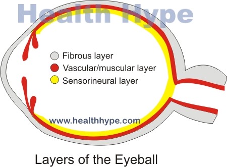
The wall of the eyeball is made up of three layers – fibrous (outer), vascular/muscular (middle) and sensorineural (inner) layers.
Diagram of the different layers of the eyeball
Outer Layer
The outer fibrous layer maintains the shape of the eyeball and protects more fragile internal structure. This layer is made up of the sclera and cornea.
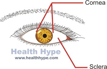
- The sclera is the firm opaque outer part of the eye commonly referred to as the “whites” of the eye. It covers most of the outer surface of the eyeball.
- The cornea is the central transparent part of the outer layer located at the center of the anterior portion (front part) of the eyeball. It is more convex (curved outwards) than the sclera and appears to protrude from the front of the eyeball.
Diagram of the sclera and cornea
The fibrous layer has a very minimal blood supply – the sclera has few blood vessels while the cornea has none. Oxygen is derived from the inner structures and oxygen in the lacrimal fluid (tear). Capillary loops located in the grayish junction between the sclera and cornea, known as the corneoscleral junction, also provides oxygen particularly to the cornea.
Middle Layer
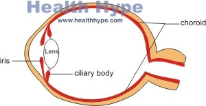
The middle vascular layer, known as the uvea is the main source of oxygen and nutrients for inner and outer linings of the eyeball. It also has muscular components that control the entry of light into the eyeball. This layer consists of the choroid, ciliary body and iris.
- The choroid is a highly vascularized layer that lies between the outer sclera and inner retina. It is a dark red color due to the concentration of blood vessels.
- The ciliary body connects the choroid to the iris and is a muscular and vascular structure lying just behind the corneoscleral junction.
- The iris is the central diaphragm at the front of the eye. It can open or close to widen or narrow the central aperture known as the pupil. In this manner, it can control the amount of light entering the eye.
Diagram of the choroid, iris and ciliary body
Inner Layer
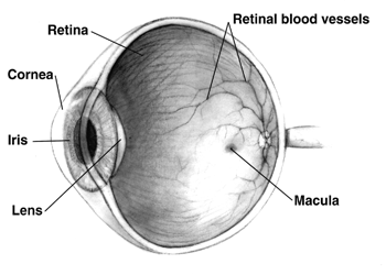
The inner sensorineural layer is known as the retina. Broadly, the retina can be divided into the neural layer that has light receptors and deciphers the light stimuli and the pigmented area that absorbs light so that it does not reflect on the neural layer and create distortion. Two other important areas are the :
- Fundus of the retina is where the light is focused. It has an area called the optic disc where the optic nerve fibers are located and this area is insensitive to light. The blood vessels of the fundus are clearly visible during opthalmic examination.
- Macula lutea is the small yellow oval spot near the optic disc. It contains the light-sensitive cones which are responsible for visual acuity. A specialized depression on the macula known as the fovea centralis is the area of the most acute vision.
Picture from Wikimedia Commons
Inside the Eyeball
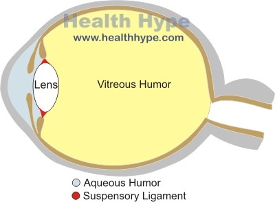
The important structures within the eye includes the lens and suspensory ligaments, and the aqeous and vitreous humor. There are two segments within the eyeball – anterior segment and posterior segment. The anterior segment is small, accounting for some 20% of the inner area of the eyeball, and lies between the cornea and anterior aspect (front) of the lens. The posterior segment which accounts for the other 80% of the inner eyeball lies between the posterior surface (back) of the lens and the retina.
Diagram of the refractive compartments and lens
The lens is an elastic, biconcave structure that lies just behind the iris and can alter shape to focus light on the retina. It is surrounded by a capsule which has fibers that keep the lens suspended in its normal position. The elastic fibers known as the zonula fibers collectively form the suspensory ligament which attaches to the ciliary body. When the ciliary muscle contracts or relaxes, the tension on the lens increases or decreases. This alters the shape of the lens so as to change the focusing of the incoming light to ensure for a more clearer image in a process known as accommodation.
The anterior segment has an anterior chamber between the cornea and iris and the posterior chamber lying between the iris and the lens. The anterior chamber is filled with a watery, nutrient rich fluid known as the aqueous humor which is produced by the ciliary processes of the ciliary body. The posterior segment is filled with a gelatinous substance known as the vitreous humor.
Orbit
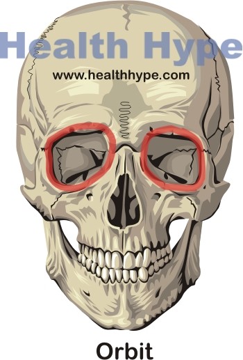
The orbit is the bony hollow region in the skull that houses the eyeball and is also referred to as the socket. It is shaped similar to a pyramid with the base facing anteriorly (front) and the apex pointing posteromedially (to the back and inner side).
Diagram of the orbit (eyeball socket) in the skull
The orbit is made up of several bones of the skull :
- Frontal bone at the top (superiorly)
- Zygomatic bone on the front and outer side (anterolaterally)
- Maxilla on the front and inner side (anteromedially)
- Lacrimal and nasal bones on the back and inner side (posteromedially)
- Ethmoid bone on the lower back (posteroinferiorly)
- Sphenoid bone on the back and outer side (posterolaterally)
The different bones extend and overlap into neighboring regions. Another prominent feature at the back of the orbital region is the optic canal through which the optic nerve passes. It is located in the sphenoid bone. The orbit is packed with orbital fat that cushions the eyeball within the socket.
Appendages
There are several accessory parts of the eye including the eyelids, lacrimal glands, extraocular muscles, nerves and blood vessels, fascia and the mucous membrane known as the conjunctiva.
Eyelids
The eyelids are folds of skin that can close to cover the eyeball. In this manner it can protect the eyeball from very bright light and injury. The eyelids are also able to ‘wipe’ away any particles that make contact with the outer layer of the eyeball and keeps the cornea moist. The inner lining of the eyelid is lined with a thin mucous membrane known as the palpebral conjunctiva which then reflects onto the eyeball where it covers the outer layer and is known as the bulbar conjunctiva.
The borders of the eyelids contain a thick flexible band of connective tissue known as the tarsi – superior tarsi (upper eyelid) and inferior tarsi (lower eyelid). Tarsal glands embedded within the tarsi secrete a fatty substance that prevents the eyelids from sticking together when closed. Eyelashes (hair) extend from the margins of the eyelids ad has ciliary glands attached to it which are sebaceous glands that secrete sebum (oil).
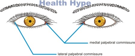
The junctions where the upper and lower eyelids meet are known as the canthi (singular ~ canthus) or commonly as the corner of the eyes or angle of the eyelids. These areas are known as the medial palpebral commissure (inner angle near the nose) or lateral palpebral commissure (outer angle).
Diagram of the outer eye
Lacrimal Glands and Associated Structures
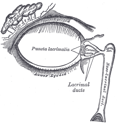
The lacrimal glands are almond-shaped glands that sit in the superolateral (upper outer) part of the orbit. It is located just under the outer part of the eyebrow. These glands produce lacrimal fluid, commonly referred to as tears, that flush out any dust particles, keep the outer lining of the eyeball moist and also contains dissolved oxygen for the cornea.
The lacrimal glands have about 10 excretory ducts that carry fluid out of the glandular substance onto the outer surface of the eyeball. It is spread by the blinking eyelids. The excess fluid drains to the medial angle of the eye, where it enters openings known as the lacrimal punctum. From here the fluid drains through the lacrimal canals into the lacrimal sac. It then drains into the nasal cavity where it is swallowed.
Picture from Wikimedia Commons
Extraocular Muscles
The six extrocular muscles move the eyelids and eyeballs.
- Levator palpebrae superioris – lifts the upper eyelid
- Superior oblique – moves eyeball to the outer side (abduct) and downwards (depresses), medial rotation
- Inferior oblique – moves eyeball to the outer side (abduct) and upwards (elevates), lateral rotation
- Superior rectus – moves eyeball to the inner side (adduct) and upwards (elevates), medial rotation
- Inferior rectus – moves eyeball to the inner side (adduct) and downwards (depresses), lateral rotation
- Medial rectus – adducts eyeball
- Lateral rectus – abducts eyeball
Nerves of the Eye
Most of the nerve supply to the eye, both motor and sensory, are via the cranial nerves.
- Optic nerve (CN II) – sense of vision
- Oculomotor (CN III), trochlear (CN IV) and abducent (CN VI) – movement of the eyeball (extraocular muscles)
- Facial nerve (CN VII) – production of lacrimal fluid
- Opthalmic nerve (CN V) – lacrimal glands, eyelids (movement), cornea (touch)

