Urinary Tract Anatomy, Location, Parts and Pictures
The organs of urinary system includes the kidneys, ureters, urinary bladder and urethra. Collectively, these organs produce urine, store it and pass it out of the body. The process, however, has far reaching consequences in the body and an integral role in maintaining homeostasis. Since the kidney controls the fluid volume and electrolyte levels in the body, it can impact on blood pressure, heart function and even influence nerve and muscle activity to some degree.
The urinary tract can be divided broadly into upper and lower parts. The kidneys and most of the upper part of the ureters comprise the upper urinary tract, while the distal parts of the ureters, urinary bladder and urethra make up the lower urinary tract. Other neighboring organs and structures which may not be part of the urinary system also impact on urinary function, in particular the genitalia, and therefore the term genitourinary system is often preferred anatomically.
Parts of the Urinary Tract
The urinary tract comprises 4 main parts :
- Kidneys (one on either side)
- Ureters (one from either kidney)
- Bladder
- Urethra
Kidneys
The kidneys are ovoid, often referred to as bean-shaped, reddish-brown organs located on either side of the upper abdomen, on the posterior abdominal wall (back). It lies behind the peritoneal cavity (retroperitoneally) and is surrounded by fat which serves to protect it. A kidney is about 10 centimeters long, 5 centimeters wide and 2.5 centimeters thick.
Each kidney has a concave medial margin (inner side) and convex lateral border (outer side). At the medcal margin is a central cleft known as the renal hilum, where blood vessels, nerves and the ureters enter or exit the kidney. The right kidney is about 2.5 centimeters lower than the left kidney due to the presence of the liver on the right side. However, the vertical position of both kidneys may vary during breathing by around 2 to 3 centimeters. Read more on kidney pain location for more information on its anatomical position.
Internally the tissue of the kidneys can be divided between the renal cortex and the renal medulla. Several cone-lobes of the medulla are surrounded by the renal cortex and these areas are the main functional zones of the kidney. It contains the nephron, which is the basic functional unit comprising the glomerulus and tubule, surrounded by kidney tissue (interstitium). Urine that is produced by the nephrons empties into the renal pelvis and then drains into the ureters.
Ureters
The ureters are narrow muscular tubes that are about 25 to 30 centimeters in length. It is continuous with the apex of the renal pelvis (kidney) an carries urine formed in the kidney to the urinary bladder. The ureters cross over from the abdominal to the pelvic cavities and run a short distance into the urinary bladder. There are three points where the ureters are slightly constricted compared to the rest of its luminal diameter :
- where it communicates with the renal pelvis (apex)
- where it crosses the pelvic inlet (lateral part of the pelvis)
- where it penetrates the wall of the urinary bladder
Urinary Bladder
The urinary bladder is hollow organ that is shaped like a tetrahedral (when empty). It resembles an inverted three-sided pyramid with a triangular base. The bladder has muscular walls that stretch to accommodate up to a maximum of 500 milliliters of urine. The wall muscle known as the detrusor muscle contracts during urination to expel the urine within the bladder.
The bladder has four surfaces – one top surface, one back surface and two sides – which are closely related to the reproductive system. In women, the uterus lies slightly above the bladder and the vagina behind it while in men, the prostate gland lies immediately below the bladder.
Inferiorly, the bladder narrows to form the bladder neck where two sphincters control the outflow of urine. The internal sphincter is made up of a circular arrangement of the detrusor muscle and is under involuntary control. The external sphincter is composed of the urogenital diaphragm which is under voluntary control. For more information on the urinary bladder, refer to the article on bladder anatomy.
Urethra
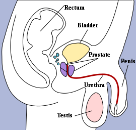
The urethra is the a narrow tube that extends from the neck of the bladder to the relative orifices in men and women where urine can be expelled out into the environment. It varies significantly in length between men and women – some 20 centimeters in men and approximately 4 centimeters in women. Apart from this difference in length, the female urethra is also narrower.
Picture from Wikimedia Commons
The male urethra, due to its length and anatomical course, can be divided into three parts :
- Prostatic urethra running through the prostate gland about 3 to 4.5 centimeters in length. A short portion that exits the bladder, just before it runs through the prostate gland, is known as the pre-prostatic urethra.
- Membranous urethra which is about 1 to 2 centimeters in length is the short portion of the urethra when it leaves the prostate gland and before it runs through the penis.
- Spongy urethra, also known as the cavernous urethra, is about 15 centimeters long and runs through the corpus spongiosum of the penis.
The external urethral orifice is the opening of the urethra into the environment. In women, it is located in the vestibule between the labia minora, while it men it is situated at the top of the penis. For more information, refer to urethra anatomy.


