Torn Knee Cartilage
A torn cartilage at the knee joint is commonly seen after a twisting injury. This generally occurs in athletic activities or road accidents. The ‘cartilage’ referred to in this colloquial term, is not the articular cartilage, which normally covers the ends of bones in a joint. It refers to a specialized cartilaginous structure present between the bones (Picture 1) at the knee joint, which is called a meniscus (pl. menisci). The menisci are highly important for stabilizing the knee as well as making it capable of bearing the weight of the body. They also help in lubricating the joint for smooth movements.
Anatomy of Menisci
There are two menisci present in each knee; one on the medial side and the other on the lateral side between the femur (thigh bone) and tibia (leg bone). Each meniscus is a C-shaped disc (Picture 2) of cartilaginous tissue which fills the gap between the femur and the tibia (Picture 3). This causes more portion of the bones to come in contact with each other and distributes the pressure evenly on the surface of the two bones.
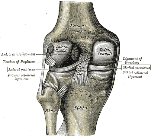 Picture 1: Menisci from behind knee
(source: Wikimedia)
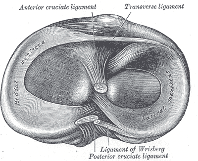
Picture 2: Menisci seen from above
(source: Wikimedia)
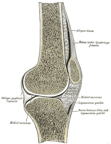 Picture 3: Cut Section of Knee showing menisci
(source: Wikimedia)
Symptoms of Torn Knee Cartilage
A torn cartilage or meniscus in the knee causes it to become swollen and painful. The pain is mainly during squatting or movements in which the knee is completely folded (flexed). The torn fragment of cartilage gets interposed between the bones and causes transient locking of the joint. A locked joint may unlock spontaneously or after some manipulation. Sometimes, the symptoms of torn meniscus are very mild and it may even go unnoticed for many years.
Diagnosis of Torn Knee Cartilage
A torn knee cartilage is not seen on X-rays, yet that is the first investigation to be done because it helps us to rule out the more obvious causes of knee pain, such as fractures.
MRI of the knee joint detects a torn cartilage and gives important information like its location and size, which helps in planning the treatment.
Arthroscopy is the investigation of choice, as the tear can be seen as well as repaired in the same sitting. It requires a highly skilled orthopedic surgeon and can be very costly. But as the joint is not entirely opened, prevents damage to other structures of the joint.
Treatment of Torn Knee Cartilage
The treatment of torn knee cartilage begins with immobilization of the joint. This is done with the help of a long knee extension brace or a plaster, which keeps the knee straight (extended). This helps the injured structures to heal and the inflammation to subside. This is done for 3-4 weeks during which the joint is gradually mobilized by a physiotherapist.
Once the swelling and pain have decreased, an MRI is done and using the information obtained from it the meniscus is stitched by an arthroscopic surgery. The surgery consists of small incisions which heal early and without much scar tissue. After surgery, again the knee is kept extended by a brace or a plaster for 2 weeks, and then followed by physical therapy.