Signs Of A Compressed Nerve In The Back
Any nerve in the body can become compressed for a number of reasons. But it is often the nerves emanating from the spine that are the most prone. These spinal nerves have to pass out between the bones of the spine (vertebrae) to different parts of the body. It is at the root, as it passes out of the vertebral column, that these nerves are more likely to become compressed. Therefore this condition is referred to as nerve root compression (radiculopathy) or simply as a pinched back nerve. Compression affects the nerve’s function which is to carry signals to and from the spinal cord.
As a result there are a number of symptoms arise – abnormal sensations when sensory nerves are compressed or muscular symptoms when the motor nerves are compressed. Back symptoms are not always present. This means that you may not even have back pain yet there are nerve root compression symptoms present at different parts of the body, like the arms and hands or legs and feet. This can be misleading because the cause of the problem is at the back, and not at the location of the symptoms.
So how do you know if a nerve in your back is compressed or not?
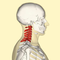
Neck (Cervical)
The first 7 vertebra lying in the neck and upper back are known as the cervical vertebrae. It has to support the head but also has to be extremely mobile to allow for neck and head mobility. Most of the nerves emanating from this part of the spinal cord supplies the shoulders, arms and hands. Therefore symptoms are evident on these areas. Read more on a pinched neck nerve.
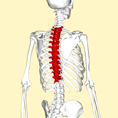
Upper and Mid Back (Thoracic)
The upper and mid back is primarily made up of the thoracic vertebrae. There are 12 thoracic vertebrae that continues from the last cervical vertebra. It corresponds to the chest area. The symptoms may overlap with cervical nerve root compression (shoulders, arms and hands) and the front of the chest as well as between the shoulder blades. Read more on pinched thoracic nerve.
Lower Back (Lumbar)
The lower back is comprised primarily of the 5 lumbar vertebrae. This continues from the last thoracic vertebra to the sacrum that makes up the back of the pelvis. The nerves emanating between these vertebrae supply the buttocks, thighs, lower leg (including the calf) and feet. Therefore nerve root compression of these nerves will cause symptoms in the lower body. Read more on pinched lower back nerve.

Common Causes
A compressed nerve root may arise for a number of different reasons. It is often associated with age-related processes and a pinched nerve is therefore seen more frequently in the elderly. But it can occur much earlier in life as well. Here are some of the common causes of a pinched spinal nerve:
- Injury to the spine like with a car accident (cervical and thoracic nerves particularly) or falling on the back.
- Herniated intervertebral discs (IV discs) which is a bulging of the spongy cushion between the spinal bones.
- Degenerative IV disc disease where there is age-related wear-and-tear of the lumbar discs in particular.
- Bone spurs which are bony outgrowths from the vertebrae that may press on the nerve roots.
- Kyphosis which is an abnormality in the curvature of the thoracic vertebrae.
- Spinal growths which may vary from tumors to abscesses.
How Do You Know If You Have A Pinched Nerve In The Back?
When a nerve root is compressed, the functioning of that specific nerve is affected. The degree to which its function is compromised varies depending on the severity of the cause and degree of nerve root compression. Remember that nerves have two type of fibers – sensory and motor. Nerve root compression can affect either of these fibers. Since many nerves are mixed (combination of sensory and motor), the functioning of both may be affected to varying degrees at the same time.
The sensory fibers carry signals from a part of the body to the spine. Some of these signals are perceived as different sensations but other signals are responsible for certain sensory information that we do not overtly perceive. Motor nerves carry signals from the spine (usually originating in the brain) to the muscles of the body. Skeletal muscles are under voluntary control and we can overtly perceive any changes in skeletal muscle activity. But it may also affect smooth muscles which are not under our conscious control but are responsible for various functions from widening or narrowing the blood vessels to movement of food and waste through bowels.
Pain, Burning and Tingling
The sensory nerves are responsible for a number of different sensations. However, when these nerves are compressed it can also cause abnormal sensations that are not due to any problem at the affected part where it is felt. But it may also be felt in the back at the site where the nerve root is compressed. The main abnormal sensations caused by nerve root compression includes pain, burning and a tingling sensation. For example, pain, burning or tingling of the feet, legs, thighs, buttocks and/or lower back may occur when the lumbar nerves are compressed. But sometimes these abnormal sensations may only be felt on the feet without any symptoms higher up the limb or at the back.
Dull Sensation and Numbness
While nerve root compression can elicit abnormal sensations, it can also reach a point where it affects normal sensory perception at an affected area. Compression may affect the transmission of incoming sensory signals. It is mainly perceived as dulling of the normal senses or even numbness where there is no sensation. For example, nerve root compression at the level of the cervical and/or thoracic vertebrae may lead to numbness in your fingers, hands or arm. Even if there is not complete numbness, you ability to perceive temperature changes, pressure (touch) and pain may be impaired to varying degrees.
Muscle Weakness and Wasting
The strength of your muscle activity is not only dependent on how big your muscles are. It is also controlled by the signals that stimulates the muscle to contract. A higher intensity signal will cause the muscle to contract more strongly. When the motor nerves are compressed, these signals may not be of the same intensity. Therefore you will perceive it as weakness. Rarely, in very severe cases the weakening is very close to paralysis. Without the constant stimulation to contract, muscles gradually start to shrink (atrophy). It is worst in paralysis but even when the signal is weak, albeit present, the muscles will gradually shrink and become smaller in bulk (muscle wasting).
Poor Motor Coordination
Coordinating activities that requires the use of muscles is controlled by the brain. But the brain is equally dependent on the feedback it receives from the part of the body that it is controlling. It is a two way process – the brain sends motor signals to a body part and alters the signaling according to the feedback it receives through sensory signals from that body part. This is how the body is able to coordinate different activities, be it kicking a ball or writing with a pen. A combination of diminished sensory feedback to the brain coupled with muscle weakness leads to poor coordination that may be seen with nerve root compression.


