Brittle Bones Tests, Scans and Other Diagnostic Investigations
Screening for osteoporosis in high risk groups like postmenopausal women, elderly men, or patients on long-term corticosteroid therapy is always advisable for early detection. Management of osteoporosis also requires recording of the baseline values of parameters for monitoring the progress of osteoporosis therapy. The diagnostic procedure becomes more complex in younger patients without any known risk factors and therefore other uncommon causes of osteoporosis also have to be investigated these individuals. The most important investigations related to early detection and management of osteoporosis are radiological investigation, laboratory tests and biomarker studies.
The traditional diagnostic tests for diagnosis of osteoporosis are radiological investigations. Radiological studies ranging from conventional radiography to complicated imaging studies may be used as an aid in diagnosis of osteoporosis. The gold standard osteoporosis diagnostic study is the bone mineral density (BMD) measurement. BMD measurement can be done with any of the imaging study options available.
Common imaging study options available for BMD estimation include :
- single/double-photon absorptiometry (SPA/DPA)
- dual-energy X-ray absorptiometry (DEXA)
- quantitative computed tomography (QCT) scanning
- magnetic resonance imaging (MRI)
EXA is the most commonly used method among the options available for measuring BMD. Best results of fracture risk assessment related to osteoporosis are obtained with imaging instruments that can give BMD from the trabecular bone than cortical bone.
Conventional X-ray
Plain radiography or x-ray is the simplest radiological investigation that is useful in diagnosis of osteoporosis. Significant changes in plain radiography are observed only after the cortical bone is involved by osteoporosis. These changes are seen only after osteoporosis is established. The thinning of the cortical bone and reduced radio-opacity of the bones are suggestive of osteoporosis in plain radiography. Plain radiography is less sensitive for diagnosis of early stage osteoporosis. Plain radiography can give early indications of complications of osteoporosis (like fractures).
Conventional radiography is generally recommended to assess the overall condition of the bones in patients with osteoporosis or those at high risk. It may be done to detect fractures in osteoporosis patients with suspected fractures. It is also useful in patients with hyperparathyroidism and osteomalacia. Plain radiography may be used in conjunction with other radiological studies like a CT or MRI. Spinal radiography is usually done in osteoporosis patients with height loss (due to vertebral height loss) or if a fracture is suspected. It is also usually done during a follow up of osteoporosis patients as fractures are common complications of osteoporosis.
Dual-energy X-ray absorptiometry (DEXA)
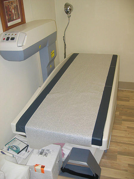
Dual-energy X-ray absoptiometry is the most common method used to estimate the bone mineral density (BMD). The wide use of DEXA is due to its lower cost, good accuracy, low radiation exposure and short scanning time. Either ends of the thigh bone, hip and the body of lumbar vertebrae are important sites for measuring bone mineral density (BMD) by DEXA. Individuals with minimal risk of osteoporotic fracture can be identified with BMD measurement of wrist using DEXA and can be excluded from further investigations in the near future. Periodic BMD measurement is the ideal way to monitor the response to therapy and to assess future risk for fracture.
BMD measurement is recommended in women past 65 years of age, postmenopausal women at risk of fracture, individuals with history of fracture due to fragile bones, patients on long term corticosteroid or anticoagulant therapy, patients diagnosed with osteoporosis and those on treatment for osteoporosis. BMD measurement for screening of osteoporosis may also be done in men past 70 years of age irrespective of risk factor status and in men past 50 years of age with risk factors. The DEXA scanners in the past were useful only in scanning in anterioposterior view while the newer DEXA scanners can scan from lateral view. The lateral view gives better view of the trabecular bone than anterioposterior view.
Results of Dexa Scans
The result of DEXA scans are given as T-scores and Z-scores. T-scores represent the comparison of the observed BMD value with that of standard control subjects. T-score system is generally applied to postmenopausal women and men past 50 years of age. The T-scores can range from less than 1 standard deviation (SD) to more than 2.5 SD below that of the mean BMD value of the adult reference population. T-scores can be categorized into four groups based on this. With reduction of each SD in measured BMD value from standard reference value, there is an increase in the fracture risk by 2-3 times.
T-score interpretation :
- T-score value within 1 SD below the average BMD reference value is ‘normal’.
- T-score value between 1 to 2.5 SD below the average BMD reference value is ‘osteopenia’ or reduced bone mass.
- T-score value less than 2.5 SD below the average BMD reference value is ‘osteoporosis’.
Severe osteoporosis is defined as T-score below 2.5 SD along with the presence of fragility fractures.
Z-scores represent comparison of measured BMD with BMD of patients of same age and gender. Z-scores are adjusted for the ethnicity is used in premenopausal women and men below the age of 50 years. Osteoporosis in these age groups are not diagnosed based on BMD measurement alone. Z-score values are categorized into measurements below the expected range for the age and measurements within the expected range for the age.
Single or dual-photon absorptiometry (SPA/DPA)
Single photon absorptiometry (SPA) measures the cortical bone density. This makes SPA less sensitive to the early-stage changes of osteoporosis that occurs in the trabecular bone. SPA may be used to measure BMD of the forearm with minimal radiation exposure. Dual-photon absorptiometry (DPA) can be used to measure BMD of the spine and upper end of femur. DPA based BMD measurements are rarely preferred as it is less accurate and time consuming.
Quantitative computed tomography scanning
Quantitative computed tomography scanning (QCT) scanning is useful only in measuring BMD of the spine. QCT scanning can measure BMD of the trabecular bone and cortical bone of the vertebral body separately. It provides the measurements in precise volumetric mineral density. This makes it the most sensitive radiological tool for diagnosis of osteoporosis by BMD measurement. However, the radiation exposure associated with QCT scanning is high. QCT scanning is more expensive and less standardized than DEXA. For these reasons QCT scanning is less preferred to measure BMD.
Quantitative ultrasound
Quantitative ultrasound may be used in assessing osteoporosis. The measurements can be done quickly without difficulty and there is no radiation exposure. It is also less expensive. The best site for quantitative ultrasound is calcaneus bone. The precision and accuracy of measurements with ultrasound is poor compared to other standard radiological investigations.
Magnetic resonance imaging (MRI)
MRI scans are useful in diagnosis of fractures. MRI scan can distinguish osteoporotic fractures from unrelated fractures.
Laboratory Studies
In addition to the changes in BMD in radiological studies, the underlying cause behind osteoporosis also needs to be investigated. Several laboratory studies are employed to determine the cause behind osteoporosis. Laboratory studies are useful in diagnosis of certain secondary types of osteoporosis. It is also useful in obtaining the baseline values of the parameters useful in assessing effectiveness of the therapy or the toxic effects of the drugs. Commonly performed laboratory investigations include routine blood studies, biochemical studies, and specific hormonal assays.
- Routine blood counts
- Serum calcium and phosphate levels
- Urinary calcium levels
- Liver function tests
- Renal function tests
- Serum electrolytes
- Vitamin D levels
- Hormone levels (thyroid, sex hormones, cortisol and parathormone)
Serum protein electrophoresis
Serum calcium levels can give early signals about the underlying cause of osteoporosis.
- Increased blood calcium is seen in hyperparathyroidism and osteoporosis associated with malignancy.
- Calcium, phosphate and vitamin D levels are useful in diagnosis of osteoporosis related to vitamin D deficiency.
- Urinary excretion of calcium in 24 hours is helpful in identifying patients with abnormal urinary excretion of calcium.
- Thyroid function tests may be performed in patients with symptoms suggestive of thyroid dysfunction.
- Sex hormones levels are useful diagnosis of osteoporosis of related to hypogonadism.
- Parathyroid hormone levels may indicate hyperparathryroidism.
- Elevated corticosteroid levels are indicative of glucocorticoid related osteoporosis.
- Multiple myeloma related bone erosions are diagnosed with help of plasma or urine protein electrophoresis.
Biomarker studies
Biomarkers have been identified in the blood and urine that can be of use in monitoring the response to treatment for osteoporosis. Biomarkers may be of bone resorption or bone formation. Increase in the markers of bone formation and bone resorption is suggestive of increased bone turnover. This pattern of biomarker levels can be seen in the early stages of postmenopausal osteoporosis. Periodic testing of biomarkers after starting therapy can give an idea about the response to the therapy.


