8 Heart Attack Tests With Pictures
If you are experiencing the symptoms of a heart attack and have to head down to the emergency room, then you know that you will be subjected to several tests to confirm the diagnosis. As scary as a heart attack is, many patients are even more scared of the diagnostic tests and surgery that may follow. But without early medical intervention, death may be knocking on your door – within minutes, hours or even days. It is only once a heart attack or other serious cardiac event is confirmed that your doctor and surgeon will look at taking you into the operating theater. First you have to undergo tests, which could even reveal that your symptoms were not related to a cardiac event that requires surgical intervention.
Types of Tests
There are several different diagnostic methods that will be employed in order to:
- Assess the functioning of the heart.
- Identify abnormalities in blood flow.
- Isolate damage to the heart muscle or valves.
- Evaluate the effects of heart problems.
These tests may measure electrical activity or chemicals from the heart and utilize sound, radiation or electromagnetic waves to visualize the heart. Some of these tests are non-invasive meaning it does not require a break in the skin for assessment, evaluation or identification of a heart problem. Other procedures are minimally invasive where an injection of a dye or introduction of a catheter into the blood vessel may be necessary through a small opening in the skin.
The two main tests that would be conducted immediately when you reach the ER on suspicion of a heart attack in an electrocardiogram (ECG) and blood tests to evaluate the cardiac enzymes. Other tests that may be conducted thereafter includes:
- Echocardiogram
- Stress ECG
- Coronary angiogram
- Cardiac imaging studies – chest X-ray, cardiac CT scan or cardiac MRI
Electrocardiogram
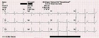
The heart has its own electrical signature that can be assessed to identify any problems. As a muscular organ, the heart is triggered to contract or relax by nerve impulses which are electrical in nature. The natural pacemaker known as the sinoatrial (SA) node generates these impulses and then it travels through the conduction system of the heart to cause a coordinated heart beat.
Electrodes placed on your chest can measure these electrical impulses occurring in your heart over a few minutes. Alternatively your doctor may advise on 24 hour monitoring with the use of a Holter monitor. Abnormalities in an ECG reading can be due to a host of cardiac problems and not only a heart attack.
Cardiac Enzymes Blood Tests
Any injury or damage to your heart muscle causes certain enzymes and proteins to leak out of the cells into the bloodstream. These cardiac enzymes or cardiac markers are useful indicators of heart problems like a heart attack. Normally these markers are present in very small quantities in the bloodstream but a surge in any marker is indicative of a problem. These markers include creatinine kinase (CK) and creatinine phosphokinase (CPK) which are enzymes, as well as proteins like troponin I (TpI) and troponin T (TpT). The levels of these markers change within hours to days after a heart attack. A blood sample is drawn and sent to a laboratory for evaluation and the results are available within hours.
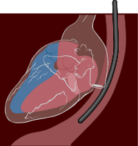
Echocardiogram
An echocardiogram is another heart test that utilizes sound waves to create images of the heart. It can allow a doctor to assess the way the heart is beating and evaluate the flow of blood through the heart. The results of an echocardiogram will indicate any abnormalities of the heart muscle, its ability to pump blood, dysfunction of the heart valves and size of the heart.
There are two types of echocardiography that may be performed. One is the transthoracic echocardiogram where the transducer is placed on the chest wall to produce images of the heart. The other is a transesophageal cardiogram where a probe containing the transducer is placed down the esophagus (food pipe).
Stress ECG
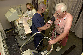
A stress ECG is an electrocardiogram where the readings are taken while a person is exercising. It is therefore also known as an exercise stress ECG. Your doctor will place the electrodes on your chest and have you walk on a treadmill. It is not usually conducted on a patient with a heart attack but is useful for diagnosing diseases that can lead to a heart attack.
In some states the electrical activity may appear to be normal at periods of rest or even with mild physical activity but once the demand on the heart increases, abnormalities may become apparent. Walking on a treadmill increases the cardiac demand and in this way heart problems like coronary artery disease (CAD) and arrhythmias become evident.
Coronary Angiogram
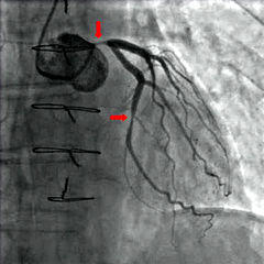
A coronary angiogram is one of the more invasive heart attack tests. By injecting a dye into the coronary arteries, the vessels that carry blood to the heart muscle, and then visualizing it on an x-ray the doctor is able to assess the degree of narrowing. Coronary artery disease is one of the main risk factors for having a heart attack and occurs when the coronary arteries become narrowed.
A catheter is inserted into the artery at your arm or thigh and directed to the coronary arteries of the heart. The dye which is visible on x-ray is then injected into the catheter while x-ray images of the heart are taken to see the coronary arteries.
Cardiac Imaging
Both an echocardiogram and coronary angiogram are cardiac imaging studies that are usually the preferred options for diagnosing a heart attack or other cardiac problems. There are also other imaging studies that may also be utilized when a heart attack is suspected. This includes a chest x-ray, cardiac CT (computed tomography) scan or cardiac MRI (magnetic resonance imaging).
Chest X-Ray
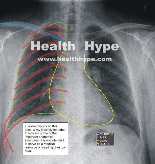
A chest x-ray is not the best imaging study to conduct in a suspected heart attack. But it is widely available and fairly inexpensive compared to other imaging studies. A chest x-ray may show a change in the size of the heart and blood vessels. Any collection of fluid in the lungs may also be evident on a chest x-ray. It cannot conclusively identify a heart attack but these findings may then prompt a doctor to conduct more specific diagnostic investigations.
Cardiac CT
A cardiac CT (computed tomography) scan utilizes a large device that moves around the body to take multiple x-ray images of the heart from different angles. By collating these images, a three dimensional (3D) image of the heart can then be formed. Sometimes a contrast dye is introduced into the coronary arteries and then a cardiac CT scan is conducted. This type of test is known as coronary CT angiography where the coronary arteries are highlighted on the 3D images of the heart.
Cardiac MRI
A cardiac MRI (magnetic resonance imaging) utilizes radio waves and a magnetic field to form 3D images of the heart from multiple angles. It is a non-invasive procedure that does not require the injection of any dyes or insertion of probes within the body. Ionizing radiation is also not used as is the case with an x-ray, coronary angiogram or cardiac CT.
A cardiac MRI can create static and moving images of the heart as well as the large blood vessels that connect to the heart. When more detailed images of the coronary arteries are needed, a contract dye known as gadolinium can be injected into the veins. An MRI is better at visualizing soft tissue while x-rays provide more detailed images of structures like bones unless a contrast dye is used in the target area.


