Chest Pain Test – Heart and Lung X-Ray, ECG Stress Tests
Every case of chest pain with one or more red flag signs or symptoms (refer to Chest Pain Diagnosis) is treated as a medical emergency. In most settings, an x-ray and ECG (electrocardiogram) are the two investigative techniques that can be conducted with immediate test results being made available.
Other tests may require periods of time for processing which is not feasible in a medical emergency. Nevertheless, if the facilities are available, the emergency room doctor may consider one or more of these chest pain tests while continuing with the appropriate treatment.
Chest pain tests are helpful in confirming a diagnosis but a skilled physician can often make a diagnosis based on clinical findings, a patient’s medical history and accurate case taking. Due to the serious nature of chest pain, many tests are conducted routinely even if serious medical disorders are not suspected by the attending doctor.
Chest X-Ray
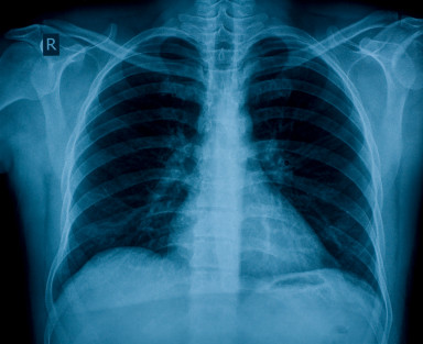
Depending on the presentation and clinical findings, a doctor may be looking for specific features on a chest x-ray. A posterioanterior (PA) and lateral x-rays will be conducted. Some of the common features that may be noted include :
- Tracheal alignment (tracheal deviation), lung segments and any signs of effusions (fluid in the lungs).
- Widening of the mediastinum, cardiac size and shape and major blood vessels shape, size and position.
- Sternal, clavicle, scapula or rib fracture.
- Change in spine curvature (lordosis and kyphosis).
- Vertebral body height and disc spaces.
- Any foreign objects or abnormal, opaque mass.
Chest X-Ray Picture. Posterioanterior View
A chest x-ray may be helpful in prompting further investigation or diagnosing the following conditions :
- Abscess, empyema
- Foreign body in the airways
- Lung cancer or tumor
- Pneumonia
- Pneumothorax
- Tuberculosis
- Thoracic aorta aneurysm and dissection
- Cardiomegaly
- Pericardial effusion
- Esophageal cancer or tumor
- Esophageal rupture
- Foreign body in the esophagus
- Gastric tumor
- Hiatal hernia
- Disc degeneration
- Fractures
- Joint dislocation
- Osteoporosis
A breast tumor may also be identified on a chest x-ray.
These are not the only conditions that may be identified with a chest x-ray. (1)
ECG, EKG (Electrocardiogram)
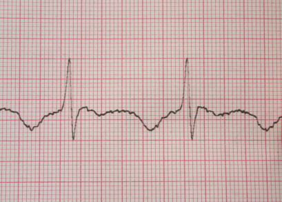
An ECG monitors the electrical activity of the heart.
This investigative technique may be useful in prompting further investigation or diagnosing the following :
- Myocardial ischemia/infarction – angina, heart attack
- Myocarditis
- Pericarditis
These are not the only conditions that may be identified with an ECG. (2)
Angiogram / Angiography
Refer to Clogged Artery Diagnosis for more information on a blocked coronary artery.
CT Angiography
A CT (computerized tomography) angiography scan uses a series of x-rays from different angles to render a 3-D view of the area. Radio-opaque contrast (dye) is injected into the bloodstream and highlights the arteries upon scanning.
It is the most effective method of assessing the blood supply to the heart. It is cheap, fast and less invasive than the angiogram mentioned below. CT angriography provides detailed images and along with an MRA (magnetic resonances angiography), it is the preferred methods of mapping coronary artery blood supply.
A coronary angiography may be conducted if any narrowing or blockage of the coronary arteries is suspected and a CT angiography is unavailable.
Coronary Angiography
Also known as an angiogram, a coronary angiography involves the injection of a special dye directly into your coronary artery. A long, flexible tube, known as a catheter, is inserted into one of the blood vessels of your arm, upper thigh or neck.
This catheter is directed to the coronary arteries where a dye is released into the blood stream and through special x-ray imaging, the flow of the dye can be monitored. This will highlight any constriction or blockage in cases of coronary artery disease. An angiography will also detect enlargement and malformation of blood vessels, even those other than the coronary arteries.
Stress Tests
In an emergency situation, a stress test will usually not be conducted. It is helpful to report any stress-induced chest pain (refer to Stress Chest Pain) as this may prompt your doctor to conduct one or more of the following stress tests as soon as possible.
Stress ECG, EKG Test
This involves an ECG test with simultaneous monitoring of the blood pressure while the patient walks (treadmill) or pedals (stationery cycle).
Stress Echocardiogram
This involves ultrasound imaging of the heart and an ECG and blood pressure monitoring is conducted simultaneously while the patient walks or pedals.
Thallium Stress Test
This test involves nuclear imaging of the blood flow through the heart muscle at rest and during activity. This procedure is not conducted as often as a stress ECG or stress echocardiogram due to the cost, need for specialized facilities and risk of complications.
Blood Tests
Full Blood Count, ESR and C-Reactive Protein
This will assist the doctor in diagnosing :
- Acute and chronic infections
- Inflammatory conditions
It is important to note that the chest pain may be a symptom of an infection or inflammation elsewhere in the body. Abnormal results may indicate a host of possible conditions and are only helpful to the doctor when the clinical findings and the results of other tests are taken into account.
For example :
- Myocarditis or pericarditis may be diagnosed based on the symptoms reported, clinical findings and the results of other tests like a chest x-ray, ECG and serum cardiac markers.
- Tuberculosis may be diagnosed based on the symptoms reported, clinical findings and the results of a chest x-ray and sputum culture.
Please note that attempting to interpret the results of diagnostic tests and thereby self-diagnose is irresponsible and dangerous. Rather leave the diagnosis to the attending doctor.
Cardiac Markers
This will assist the doctor in identifying inflammation, ischemia and infarction of the heart muscle.
Serum cardiac markers will be useful for diagnosing :
- Myocardial infarction (MI or heart attack)
- Pericarditis
- Myocarditis
Other Blood Tests
A serum amylase and lipase may be requested if pancreatitis is suspected.
H.pylori antibody test may be requested if a peptic ulcer or gastritis is suspected.
Other Investigations
Based on the symptoms that are present, clinical findings, case history and/or the results of other tests, the doctor may request further tests to diagnose the cause of the chest pain. These investigative techniques may include :
- MRI (magnetic resonance imaging)
- Ultrasound
- Endoscopy
- Barium swallow
References
- Chest X-Ray. Medline Plus
- ECG. Medline Plus


