Fluid in the Lungs (Pulmonary Edema) Causes and Treatment
Fluid in the lungs specifically refers to a condition known as pulmonary edema. However, the term may sometimes be confused with other conditions like fluid outside or aroun the lungs which is pleural effusion. Both causes characteristic symptoms, like a bubbling sound in the lungs (rales) when breathing. The term “fluid in the lungs” is also used to refer to mucus inside the lungs. Mucus or phlegm is a thick, sticky secretion while “lung water” is a thin fluid. Other fluid accumulation may be the result of blood or pus.
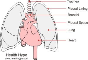
The lungs are located in the thorax (chest) and lies on either side of the heart. Air travels through the air passages, which includes the nose, pharynx (throat), trachea (air pipe) and bronchi. The lung tissue is made of small air sacs, known as alveoli, which is thin and surrounded by blood capillaries.

The structure of the respiratory system allows for an exchange of gases so that essential oxygen is taken into the body and waste products, along with gases, are excreted through the exhaled air. The lung is enclosed in an air tight pleural cavity, with a small pleural space separating the lung from the chest wall. This cavity is lined by the pleural lining, which also produces a little pleural fluid to reduce friction between the chest wall and lungs during breathing.
What is pulmonary edema?
Pulmonary edemia is the accumulation of fluid in the lung spaces. This fluids can be tissue fluid, plasma (from the blood), blood or mucus (phlegm), which is produced by the lining of the respiratory tract. The respiratory tract is lined with a mucus membrane, which is a specialized tissue that produce smucus.
This mucus lubricates the lining, which may dry out due to the air moving in and out of the passages, as well as to trap any dust or microorganisms in the air. However in certain conditions, the mucus membranes of the air passages may produce excessive amounts of mucus and this may slowly ‘sink’ lower down the air passages until it settles in the lungs.
The cough reflex or even spontaneous coughing will usually propel most mucus out through the mouth (sputum), however in cases of excessive mucus production, obstructive airway disease or diminished coughing, the mucus build up will quickly settle in the lungs.
Water in the Lungs
“Lung water” or water in the lungs usually results from interstitial fluid or blood plasma and may be an indication of a more serious underlying disorder, usually cardiovascular conditions. This fluid inside the lung is known as pulmonary edema and may be accompanied by a shortness of breath or difficulty breathing (dyspnea), a feeling of suffocation, anxiety and restlessness. Abnormal breathing sounds are also present, particularly crackling. Pulmonary edema may be considered a medical emergency and immediate medical intervention is required.

Blood inside the Lungs
Blood may also fill inside the lungs but this usually occurs as a result of severe trauma and the cause is clearly evident, like in a gunshot or stab wound. In most trauma cases where blood can fill into the lungs, the lungs collapse and the blood accumulates around the lungs in the pleural space (hemothorax). Infections like tuberculosis (TB) or lung cancer may also cause blood accumulation inside the lungs. Depending on the severity of the trauma, blood inside the lungs will cause drowning and requires immediate medical attention. Pus may also occur inside the lung due to a lung abscess and also requires immediate urgent medical attention.
Causes of Fluid Inside the Lungs
- Bronchitis is the most common cause of mucus in the lungs and is often characterized by persistent coughing. This respiratory condition may develop after the common ‘cold’ or flu (seasonal influenza) , often a s a result of a secondary bacterial infection but may also be chronic and non-infectious as in the case of smokers.
- Respiratory infections may cause hypersecretion of mucus in the respiratory tract and/or pulmonary edema and this includes viral (example – H1N1 swine flu, SARS – Sudden Acute Respiratory Syndrome, MERS – Middle East Respiratory Syndrome), bacteria (example – tuberculosis, streptococcal or pneumococcal pneumonia), fungi (example – histoplasmosis, aspergillosis, candidiasis) and parasitic (example – toxoplasmosis) infectious agents.
- Non-infections pneumonia may also result in “lung water” or fluid with a thinner viscosity. This may arise only at the affected lobe of the lung due to inflammation of the lung tissue. Pneumonia is not only caused by infections but may be due to gastric contents that are aspirated from the stomach into the lungs as is the case in aspiration pneumonia.
- Respiratory allergies often result in increased mucus production, however, in certain acute cases, there may be pulmonary edema. Post nasal drip may often lead to mucus collection within the lungs and allergies may cause also inflammation of the bronchioles and mucus in the lungs in asthma.
- Near drowning results in fluid in the lungs and even if all the fluid is drained from the lungs, it is important to monitor the patient in hospital to prevent dry drowning.
- Many cardiovascular conditions will possibly lead to pulmonary edema and this includes hypertension (high blood pressure), myocardial infarction (heart attack), heart valve disease or cardiomyopathy (damaged heart muscle).
- Hypoalbuminemia may occur due to kidney failure, liver disorders, nutritional deficiencies or protein losing enteropathy.
- Renal failure may cause pulmonary edema as the kidneys are unable to filter out toxins in the blood.
- Smoke inhalation may cause severe inflammation of the lung tissue, which results in fluid accumulation in the lungs.
- Lymphatic insufficiency result in inadequate drainage of lymph fluid.
- Drug side effects may result in pulmonary edema and this includes OTC (over-the-counter) or prescription drugs, narcotics or anesthetics. This may also occur after usage of the drug, when the effects of the drug appear to have worn off.
- Inhaled, ingested or injected toxins or poisons may increase the permeability of capillary walls, thereby leading to pulmonary edema. Some toxins may also increase mucus production in the lining of the lungs.
- Autoimmune diseases like pulmonary sarcoidosis may cause fluid in the lungs due to the inflammation of the lung tissue.
- Shortage of oxygen due to high altitudes, COPD (chronic obstructive pulmonary disease) and suffocation may result in pulmonary edema.
Fluid Outside the Lungs
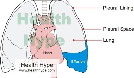
Pleural effusion is when fluid accumulates around the lung, in the pleural space. Blood (hemothorax), fatty lymphatic fluid (chylothorax) or pus (empyema) may also fill the pleural space although this occurs less frequently. Any fluid accumulation around the lungs should be taken seriously and requires immediate medical attention. The fluid accumulation around the lungs compress the lung and this prevents normal respiration, which results in inadequate gas exchange. The types and causes of pleural effusions are discussed in detail under fluid around the lungs.

Some Causes of Fluid Around the Lungs
- Congestive cardiac failure is one of the most common causes of a pleural effusion. This fluid is more thicker (transudative) due to protein that is ‘forced’ out of the blood vessels and into the pleural space.
- An exudative effusion is a watery fluid accumulation due to inflammation, caused by lung cancer like pleural mesothelioma, infections like TB or pneumonia, lung disease like asbestosis or drug reactions.
- A hemothorax may be a result of a trauma or rupture of large blood vessels in the case of an aortic aneurysm although the latter causing a pleural effusion is uncommon.
- An empyema is the accumulation of pus within the pleural space often due to a lung abscess.
- A chylothorax is the accumulation of lymphatic fluid, which has a high concentration of fat, and may occur in certain cancers like lymphoma.
- Some of the causes of fluid accumulation inside the lungs may also cause a pleural effusion, including kidney failure and liver disease.
Diagnosis of Fluid in the Lungs
Upon physical examination, your doctor will be able to identify abnormal sounds like bubbling or crackling (rales) with a stethoscope upon respiration. A wheezing sound (stridor) may be clearly audible as well when exhaling. Percussion is a tapping motion conducted against the chest wall and will assist your doctor with identifying areas of the lung that may be affected.
Typically fluid accumulation causes a dull sound compared to the normal hollow sound of the air filled lung. Based on clinical findings and other signs and symptoms, your doctor may request further diagnostic investigations, which may include the following procedures.
- A chest x-ray is one of the main diagnostic investigations conducted to identify the severity and area that is affected. For further imaging, a thoracic CT scan or chest ultrasound may be conducted.
- Due to the incidence of cardiovascular disorders related to fluid in the lungs, your doctor may conduct an ECG (electrocardiography), ultrasound of the heart (echocardiography) and other cardiac investigations.
- Fluid may be aspirated from the pleural space, which is known as thoracentesis, but this has to be carefully done to prevent a pneumothorax (accumulation of air in the pleural space). A pleural fluid analysis is then conducted to identify the type of exudate or any microorganisms.
- A sputum culture may be necessary to identify the cause of infection.
- A range of blood tests may be requested by your doctor to verify kidney and liver function, proper gas exchange and heart disorders.
Treatment of Pulmonary Edema
Treatment is dependent on the cause of the fluid in the lungs. Some of the treatment options may include :
- Antibiotics, antivirals or antifungals may be necessary in the case of an infection.
- Diuretics assist with passing out additional fluid but should be used cautiously in the case of cardiac diseases.
- Antihistamines may be necessary in allergic reactions and this may need to be continued on a chronic basis to prevent exacerbations.
- Corticosteroids may be useful for controlling inflammation and mucus production, as in asthma, and this may be used long term to prevent acute attacks.
- Chest drainage with a tube may be necessary for an empyema or a therapeutic thoracentesis may be required for a pleural effusion.
- Anti-hypertensive drugs may be administered in cases of high blood pressure.
- Oxygen is administered in severe cases of fluid in the lungs where proper gas exchange is impaired. While this does not immediately treat the cause of fluid in the lungs, except in a shortage of oxygen, it assists with adequate gas exchange.
- Physical therapy may be necessary to assist with mucus drainage.
References
- Pulmonary Edema. Merck
- Chylothorax. Medscape
Last updated on August 19, 2018.


